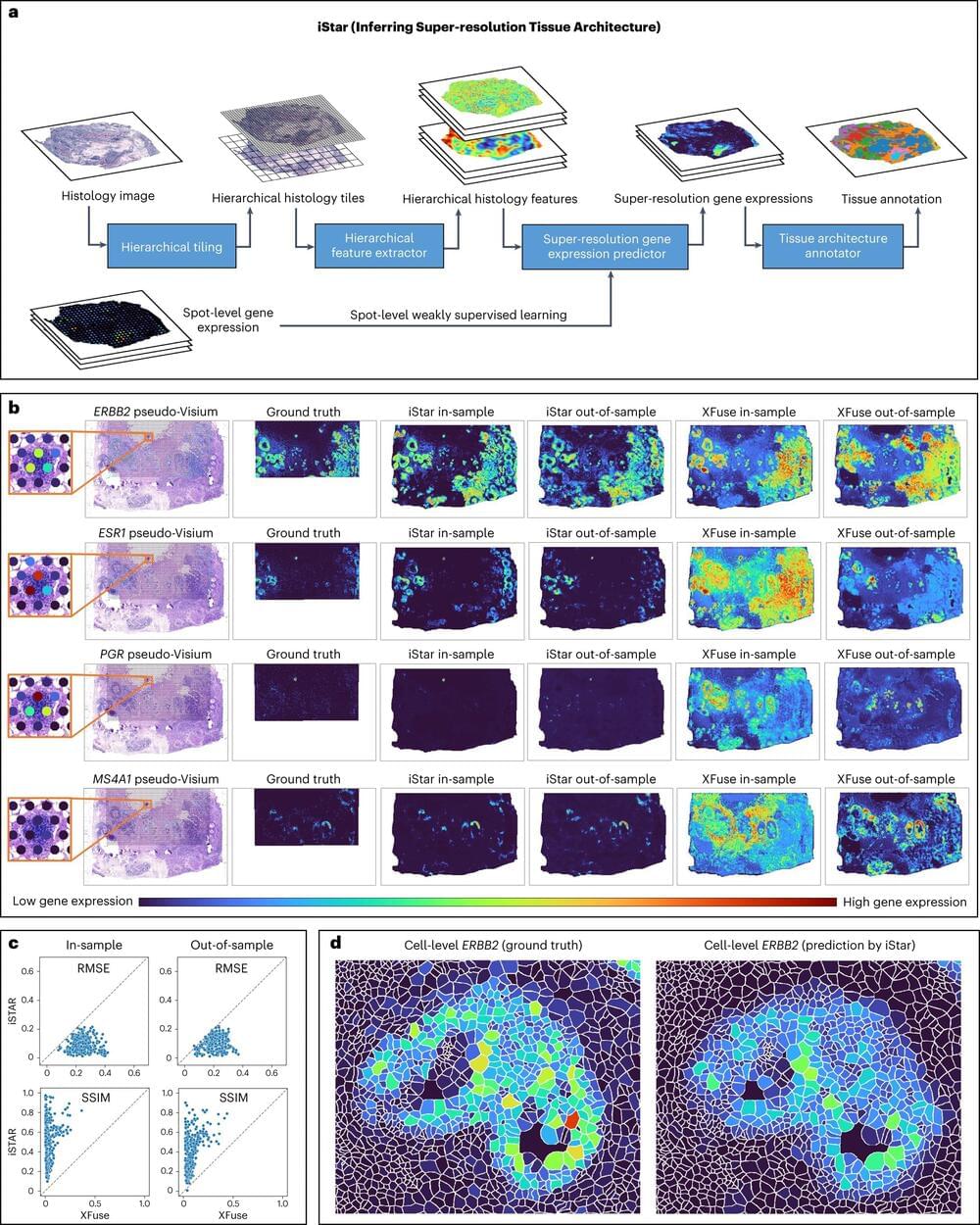
A new artificial intelligence tool that interprets medical images with unprecedented clarity does so in a way that could allow time-strapped clinicians to dedicate their attention to critical aspects of disease diagnosis and image interpretation.
The tool, called iStar (Inferring Super-Resolution Tissue Architecture), was developed by researchers at the Perelman School of Medicine at the University of Pennsylvania, who believe they can help clinicians diagnose and better treat cancers that might otherwise go undetected.
The imaging technique provides both highly detailed views of individual cells and a broader look at the full spectrum of how people’s genes operate, which would allow doctors and researchers to see cancer cells that might otherwise have been virtually invisible. This tool can be used to determine whether safe margins were achieved through cancer surgeries and automatically provide annotation for microscopic images, paving the way for molecular disease diagnosis at that level.


Comments are closed.