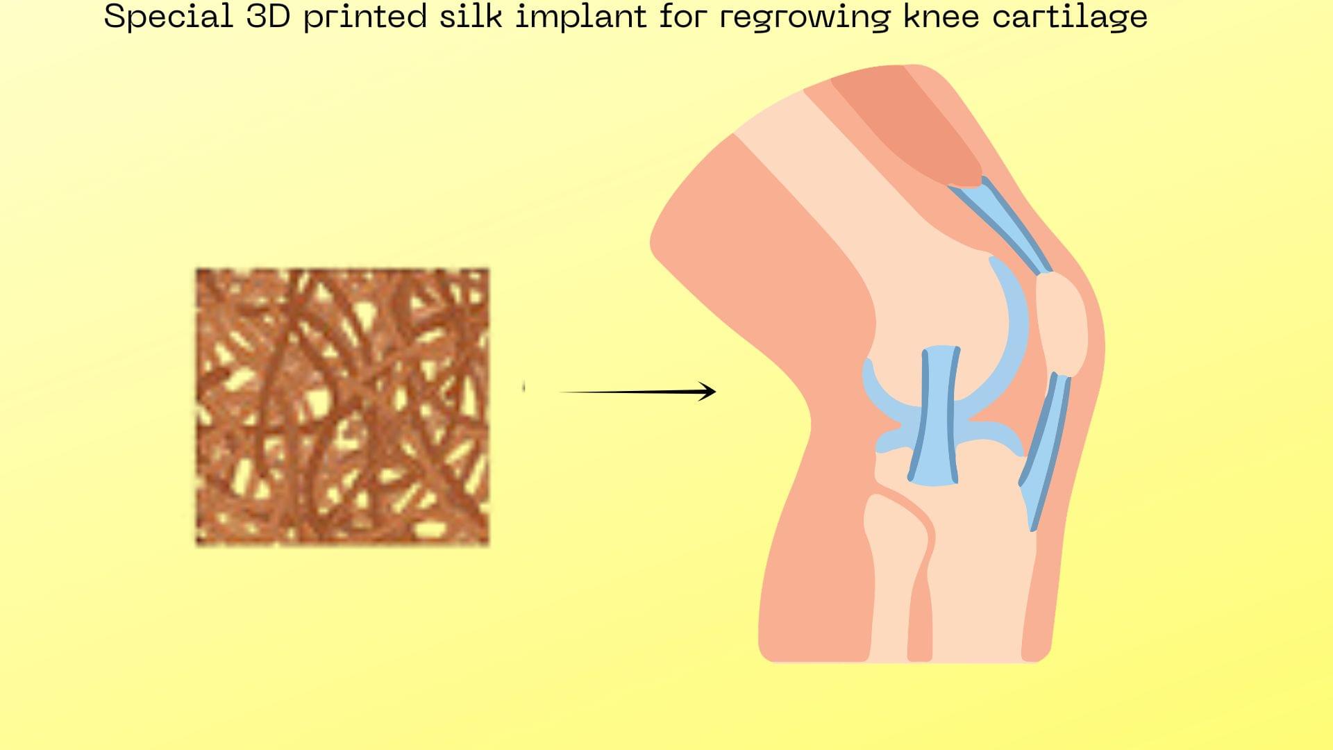
Researchers have successfully regenerated healthy knee cartilage in an osteoarthritic joint using a 3D-printed silk implant laced with a targeted drug, offering hope for a permanent cure to the debilitating joint disease.

A passive 3D-printed panel could redirect wireless signals around corners without electronics or power. The metacrystal design can handle multiple incoming waves and different frequency bands, offering a lower-cost option for hard-to-reach indoor spaces.
Basements, tunnels, large buildings—a weak Wi-Fi or mobile signal in these hard-to-reach places is frustrating. The usual solution is to add more electronics like routers, repeaters and base stations. Yet, as we move towards a 6G mobile network, this kind of complex infrastructure can be unsustainable and prohibitively expensive. Higher-frequency channels of 6G communications aim to provide vastly more data bandwidth than the current 5G, but those channels are more easily blocked by walls, people and other obstacles.
To tackle this, researchers at Aalto University have developed a new solution in the form of metacrystals: passive, 3D-printed smart panels that can shape wireless signals without electronics, a power supply or active tuning. The paper, “Metacrystals: Inversely-designed 3D-printed intelligent panels for 6G communications” is published in Nature Communications.
“When a room is too dark, you can bring in more lamps—or use simple mirrors to guide the already available light. This is what these metacrystals do, but with radio waves,” explains doctoral researcher Mahdi Asgari. “Unlike previously proposed single-layer intelligent surfaces, these volumetric metacrystals can be designed to control multiple incoming signals or frequency bands independently—a key requirement for realistic wireless communication.”
Year 2024
Caption :
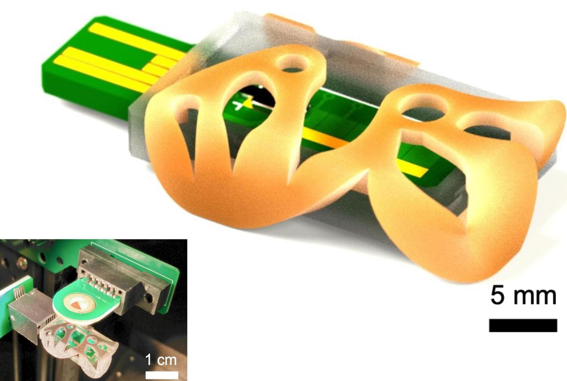
MIT researchers have 3D printed a miniature ionizer, which is a key component of a mass spectrometer. The new miniature ionizer could someday enable an affordable, in-home mass spectrometer for health monitoring. Pictured are parts of the new device, including a green printed circuit board (PCB) with orange casing on top. Under the casing is a black rectangle where the electrospray emitter is located.
Hi all, you should check out this human enhancement game created by Anton Kulaga and his team! You’ll learn about various genetic factors with potential for increasing human capabilities. The content is all rooted in scientific literature! Once you design a character, you can also get a 3D printed shape sent to you which is influenced by your choices.
Build your post-human character from real genes — tardigrade radiation shields, naked-mole-rat cancer resistance, Greenland shark longevity — backed by scientific evidence tiers and real citations. Spend enhancement credits and 3D-print the result.
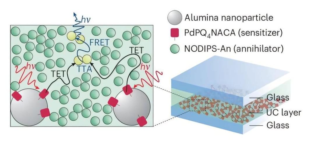
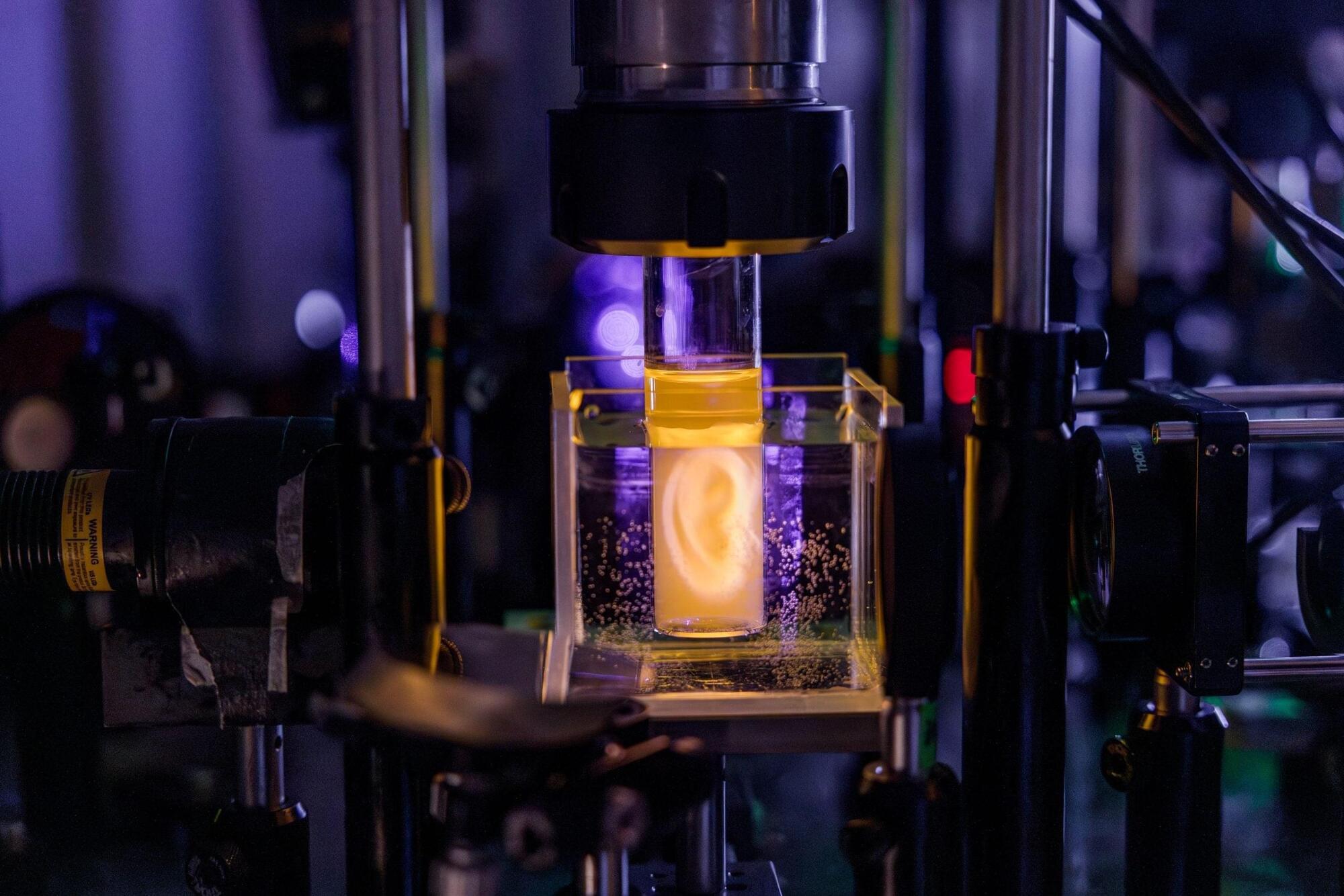
Researchers at UNSW Sydney have developed a nanoscale device that converts low-energy infrared and red light into higher-energy visible light, a breakthrough that could eventually improve solar panels, sensing technologies, and advanced manufacturing systems.
Published in Nature Photonics, the research addresses a longstanding problem in photonics: how to stop energy from being lost before it can be used.
That mechanism allowed the device to achieve photon conversion efficiencies of 8.2%, among the strongest reported for this type of architecture.
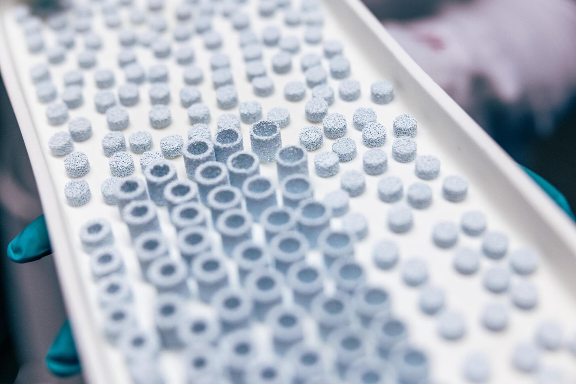
Researchers at Tampere University, Finland, have developed a groundbreaking 3D-printed ceramic implant material that closely mimics real human bone. The findings advance the development of personalized bone regeneration and may lead to more effective and accessible treatments for bone defects.
The research article, titled “Biomimetic bone calcium phosphate-based scaffolds fabricated via ceramic vat photopolymerization: Effect of porosity, sintering temperature, mineralogical phases and trace elements on the osteogenic potential,” was published in Materials Today Bio.
Bone grafting is the second most common tissue transplantation procedure worldwide, with more than 2 million operations performed annually. Current treatments often rely on bone taken from the patient or a donor, approaches that are limited in availability and may involve additional surgery, lengthy recovery times and complications. As populations age, the need for safer and more effective alternatives is growing rapidly.
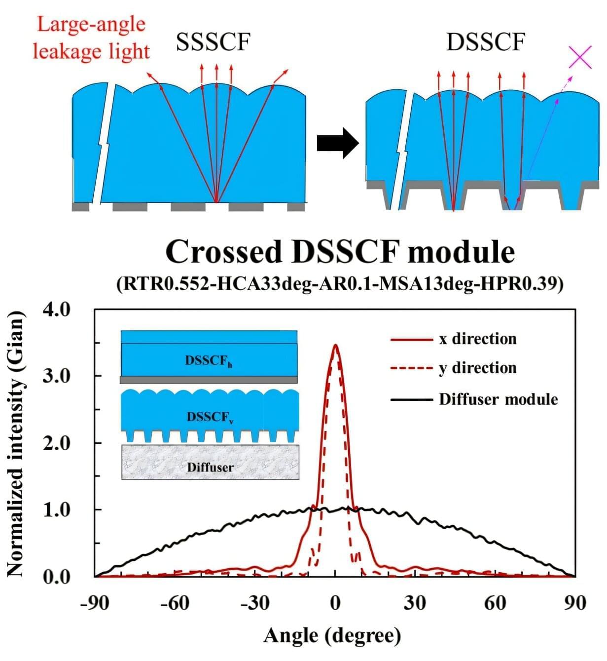
Researchers have developed an ultra-thin optical film that improves the quality of the light used in LCD resin-based 3D printers. The advance helps ensure that tiny details are reproduced with precision, which could make it possible to 3D-print medical-grade or industrial-grade products at a lower cost.
Resin-based 3D printing, or vat photopolymerization, uses short-wavelength light to project patterns onto liquid photosensitive resin. Although this additive manufacturing approach enables highly detailed, smooth parts, some low-cost systems rely on LCD backlights that can reduce printing accuracy.
“LCD-based liquid 3D printing suffers from surface roughness or dimensional inaccuracies due to improper light angular distribution from the backlight systems used,” said research team leader Ding-Zheng Lin from National Taiwan University of Science and Technology. “Our goal was to fix these problems without increasing equipment size, thereby elevating print performance to professional grade.”