Unlike accidental cell death, some cells can actively decide to die through a controlled process. This is called programmed cell death and can occur in different forms, including apoptosis and necroptosis. Cells use this process when they are damaged, stressed, becoming cancerous, or infected by harmful microbes. This self-destruction mechanism helps to protect the body, but it is also involved in many diseases, such as infections, inflammatory conditions and cancer.
A major problem in cancer is that some tumors and cancer cells learn how to avoid apoptosis, allowing them to survive when they should die. This resistance can make cancer treatments less effective, especially in advanced or spreading (metastatic) cancers.
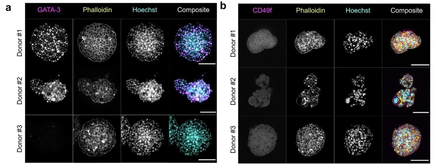
A research team led by Prof. Dr. Sjoerd van Wijk, Professor for Cell Biology at the Institute for Physiology and Cell Biology of the University of Veterinary Medicine (TiHo), and Dr. Francesco Pampaloni of the Goethe University Frankfurt, have studied a type of programmed cell death called necroptosis in advanced breast cancer. The scientists used patient-derived organoids, which are tiny 3D mini-tumors grown in the lab from real patients’ cancer cells. These mini tumors closely resemble the original cancer, making them useful for testing treatments and cell biology experiments.

