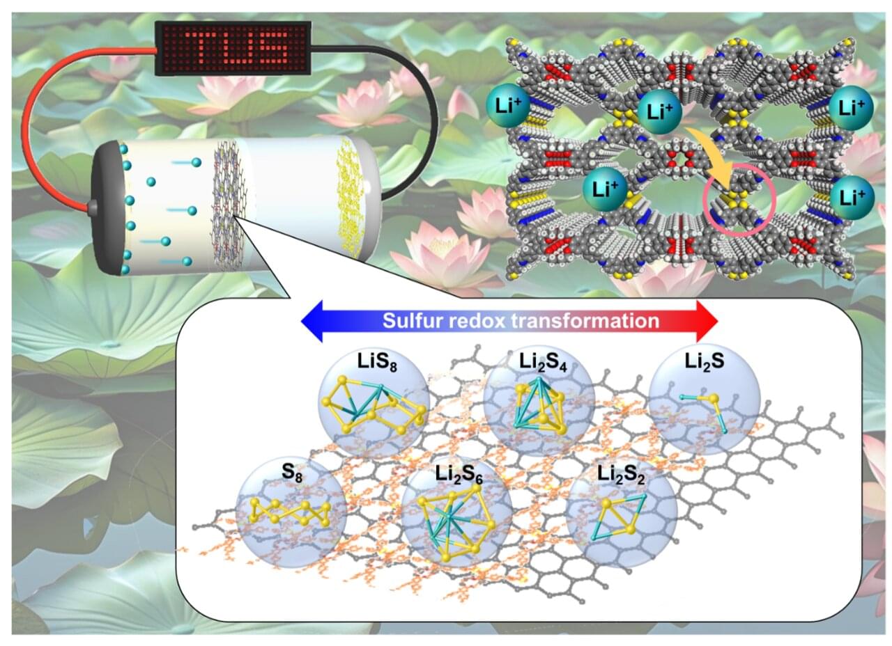
Toggle light / dark theme

Consequences of the Novel ALS-Associated KIF5A Variant c.2993-6C

Regulation and activation of UvrD-family DNA helicases/ translocases.

For the past few decades, the active form of superfamily 1A (SF1A) UvrDfamily helicases has been controversial due to the absence of structures of the active dimeric form of these enzymes.

A key interaction in the monomeric structures is between a regulatory domain (2B) and duplex DNA that was proposed to facilitate DNA unwinding but is likely inhibitory.

However, recent cryo-EM structures show that Mycobacterium tuberculosis UvrD1 forms a covalent dimer, with dimerization occurring between the 2B domains of each subunit, resulting in major reorientations of the 2B domains that prevent the 2B–DNA interaction, thus relieving its inhibitory effect.

The same dimerization interface is used in Escherichia coli UvrD dimers, suggesting that this is a general mechanism to activate most SF1A helicases.

Due to these insights, textbook descriptions of helicase mechanisms based on the monomeric structures require re-evaluation. sciencenewshighlights ScienceMission https://sciencemission.com/conundrum-resolved


AI rebuilds molecules from exploding fragments

Researchers at the Department of Energy’s SLAC National Accelerator Laboratory and collaborating institutions recently built a generative AI model that can recreate molecular structures from the movement of the molecule’s ions after they are blasted apart by X-rays, a technique called Coulomb explosion imaging.

The research, published in Nature Communications, is an important step toward being able to take snapshots of molecules during chemical reactions—an advance that could have important impacts in medicine and industry. The machine learning model closely predicted the geometries of a range of different molecules made of less than ten atoms, paving the way for applying the technique to larger molecules.

“We were pretty excited about this,” said Xiang Li, an associate scientist at SLAC’s Linac Coherent Light Source (LCLS) and lead author of the study. “It is the first AI model built for molecular structure reconstruction from Coulomb explosion imaging.”

Most mass spectrometers can process just a few molecules at once: Reengineered prototype does a billion simultaneously

Mass spectrometry is already a powerful tool for determining what kind and how many molecules are present in a given sample. But most instruments still analyze their molecules one or just a few at a time, an approach that is inefficient and costly, and in which rare, but significant molecules can easily fall between the cracks.

A more powerful version of the technology could one day allow scientists to read the full molecular contents of a single cell, track thousands of chemical reactions at once, and ultimately accelerate efforts like drug development.

Now, a new study describes the first big step in that direction by producing a prototype, dubbed MultiQ-IT, that’s capable of handling vast numbers of molecules at once. The findings, published in the journal Science Advances, offer a blueprint for faster, more sensitive instruments that could position mass spectrometry for the kind of transformation that reshaped genomics and computing.

New DNA base editor minimizes bystander edits while maintaining high efficiency

The trajectory of base editing has been remarkable, progressing from the laboratory to patient care, treating debilitating or terminal illnesses, in less than a decade. A type of gene editing that makes chemical changes to our DNA, base editing was developed by Alexis Komor, associate professor in the Department of Biochemistry and Molecular Biophysics at the University of California San Diego.

For all of base editing’s success, it is still a relatively new technology, and researchers like Komor are working to improve its efficiency, while lowering the incidence of unwanted edits. One type of unwanted edit is called a bystander edit. This occurs when a base editor not only edits the desired nucleobase, but also edits surrounding bases as well. Komor’s lab has developed a way to minimize bystander edits. This work appears in Nature Biotechnology.

The Effect of Exogenous Acid Identity on Iron Tetraphenylporphyrin-Catalyzed CO2 ReductionClick to copy article linkArticle link copied!

‘The Effect of Exogenous Acid Identity on Iron Tetraphenylporphyrin-Catalyzed CO2 Reduction’ from Inorganic Chemistry is currently free to read as an ACSEditorsChoice.

📖 Read the article.


Iron tetraphenylporphyrin (FeTPP) is a privileged electrocatalyst for the 2e–/2H+ reduction of CO2 to CO. FeTPP-catalyzed CO2 reduction typically employs phenol as an exogenous acid to promote the rate-limiting proton-coupled electron transfer. Beyond the observation that catalytic rates increase with decreasing pKa, the effects of acid identity on reaction kinetics are largely unexplored. Herein, we report rates of FeTPP-catalyzed CO2 reduction with structurally diverse O–H, N–H, and C–H acids. While many of these acids follow the expected Brønsted relationship, there are several notable exceptions: the fluorinated alcohols hexafluoroisopropanol (log(kcat) = 4.54) and 2,2,2-trifluoroethanol (log(kcat) = 3.55)─and the N–H acid imidazole (log(kcat) = 4.41)─display catalytic rates that are several times greater than rates obtained with similarly acidic phenols. Amides with pKas 19 (in dimethyl sulfoxide) display similar activity as comparably acidic O–H acids, while rates obtained with less acidic amides are ∼2 orders of magnitude slower than O–H donors of similar pKa. Each C–H acid affords poor activity. An Eyring analysis suggests that acids enforcing less ordered transition states afford superior kinetics. This study reveals that acid pKa is only one relevant parameter for altering catalytic rates, and judicious selection of the acid is crucial for enhancing catalytic rates.

Study maps gene activity linked to neurotransmission in living brains

Researchers have identified a distinct and reproducible gene expression program associated with neurotransmission in the living human brain, offering unprecedented insight into the molecular mechanisms that support human cognition, emotion, and behavior. The findings were published February 19 in Molecular Psychiatry.

Neurotransmission-the electrical and chemical signaling between neurons-is fundamental to all brain function. Until now, most gene expression studies of the human brain have relied on postmortem tissue, limiting scientists’ ability to understand which genes are actively involved in real-time neuronal communication.

In this study, investigators integrated gene expression profiling from the prefrontal cortex with direct intracranial measures of neurotransmission collected from the brains of more than 100 individuals as they underwent neurosurgical procedures. By combining molecular data with real-time physiological recordings, the team identified a coordinated set of genes whose activity tracks with neuronal signaling-a transcriptional program associated with neurotransmission.

AI model predicts chemical effects on gene expression, speeding drug discovery

Inside a diseased cell, the genes are in chaos. Some are receiving signals to overproduce a protein. Others are reducing activity to abnormal levels. Up is down and down is up. The right molecule could restore order, reversing dysregulation in specific genes. But finding the ideal compound could require examining millions of chemicals for their influence on hundreds or thousands of genes.

An MSU-led team of researchers has demonstrated a better way. Using machine learning trained on enormous amounts of published data, they were able to predict how chemicals will influence gene expression, based solely on the structure of the chemical.

Their study, recently published in the journal Cell, has discovered compounds that are promising for treatment of two difficult diseases: the most aggressive form of liver cancer and a chronic lung disease with no curative options.

Deep-learning-based de novo discovery and design of therapeutics that reverse disease-associated transcriptional phenotypes

Bulk and single-cell transcriptomics are widely used to characterize diseases and cellular states but remain underexplored for de novo drug discovery. Here, we present a strategy to screen and optimize compounds by matching disease transcriptomic profiles with compound-induced transcriptomic features predicted from chemical structures using a deep-learning model.

Gene expression program linked to neurotransmission in the living human brain identified

Researchers have identified a distinct and reproducible gene expression program associated with neurotransmission in the living human brain, offering unprecedented insight into the molecular mechanisms that support human cognition, emotion, and behavior. The findings were published in Molecular Psychiatry.

Neurotransmission—the electrical and chemical signaling between neurons—is fundamental to all brain function. Until now, most gene expression studies of the human brain have relied on postmortem tissue, limiting scientists’ ability to understand which genes are actively involved in real-time neuronal communication.

In this study, investigators integrated gene expression profiling from the prefrontal cortex with direct intracranial measures of neurotransmission collected from the brains of more than 100 individuals as they underwent neurosurgical procedures. By combining molecular data with real-time physiological recordings, the team identified a coordinated set of genes whose activity tracks with neuronal signaling—a transcriptional program associated with neurotransmission.

Laser-assisted electron scattering seen with circularly polarized light for the first time

Researchers from Tokyo Metropolitan University have succeeded in detecting laser-assisted electron scattering (LAES) using circularly polarized light for the first time. The use of circularly polarized light promises valuable insights into how atomic scale “helicity” impacts how electrons interact with matter and light.

Using synchronized femtosecond laser pulses and electron pulses directed at argon atoms, they succeeded in detecting a LAES signal showing excellent agreement with theory. The findings are published in The Journal of Chemical Physics.

LAES is a cutting-edge tool for understanding how electrons interact with matter under the influence of strong fields. When electrons are fired at atoms or molecules, they are scattered in all directions; the presence of strong light can change the way in which the scattering takes place due to an exchange of energy with the surrounding light field.

/* */