Toggle light / dark theme

Dr. Matthew MacDougall: Neuralink & Technologies to Enhance Human Brains | Huberman Lab Podcast

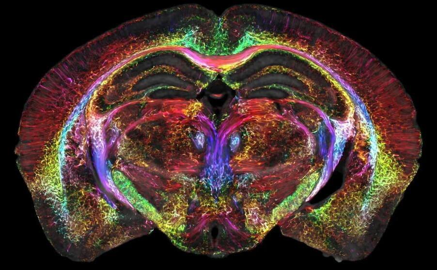
In this episode, my guest is Matthew MacDougall, MD, the head neurosurgeon at Neuralink. Dr. MacDougall trained at the University of California, San Diego and Stanford University School of Medicine and is a world expert in brain stimulation, repair and augmentation. He explains Neuralink’s mission and projects to develop and use neural implant technologies and robotics to 1) restore normal movement to paralyzed patients and those with neurodegeneration-based movement disorders (e.g., Parkinson’s, Huntington’s Disease) and to repair malfunctions of deep brain circuitry (e.g., those involved in addiction). He also discusses Neuralink’s efforts to create novel brain-machine interfaces (BMI) that enhance human learning, cognition and communication as a means to accelerate human progress. Dr. MacDougall also explains other uses of bio-integrated machines in daily life; for instance, he implanted himself with a radio chip into his hand that allows him to open specific doors, collect and store data and communicate with machines and other objects in unique ways. Listeners will learn about brain health and function through the lens of neurosurgery, neurotechnology, clinical medicine and Neuralink’s bold and unique mission. Anyone interested in how the brain works and can be made to work better ought to derive value from this discussion.

#HubermanLab #Neuroscience.

Thank you to our sponsors.
AG1 (Athletic Greens): https://athleticgreens.com/huberman.
HVMN: https://hvmn.com/huberman.
Levels: https://levels.link/huberman.
Thesis: https://takethesis.com/huberman.
InsideTracker: https://insidetracker.com/huberman.

Supplements from Momentous.
https://www.livemomentous.com/huberman.

Huberman Lab Social & Website.
Instagram: https://www.instagram.com/hubermanlab.
Twitter: https://twitter.com/hubermanlab.
Facebook: https://www.facebook.com/hubermanlab.
LinkedIn: https://www.linkedin.com/in/andrew-huberman.
Website: https://hubermanlab.com.
Newsletter: https://hubermanlab.com/neural-network.

Dr. Matthew MacDougall.

Brain images just got 64 million times sharper

Magnetic resonance imaging (MRI) is how we visualize soft, watery tissue that is hard to image with X-rays. But while an MRI provides good enough resolution to spot a brain tumor, it needs to be a lot sharper to visualize microscopic details within the brain that reveal its organization.

In a decades-long technical tour de force led by Duke’s Center for In Vivo Microscopy with colleagues at the University of Tennessee Health Science Center, University of Pennsylvania, University of Pittsburgh and Indiana University, researchers took up the gauntlet and improved the resolution of MRI leading to the sharpest images ever captured of a mouse .

Coinciding with the 50th anniversary of the first MRI, the researchers generated scans of a that are dramatically crisper than a typical clinical MRI for humans, the scientific equivalent of going from a pixelated 8-bit graphic to the hyper-realistic detail of a Chuck Close painting.

A new breakthrough in Alzheimer’s disease research: Visualizing reactive astrocyte-neuron interaction

Recently, a team of South Korean scientists led by Director C. Justin Lee of the Center for Cognition and Sociality within the Institute for Basic Science made a discovery that could revolutionize both the diagnosis and treatment of Alzheimer’s Disease. The group demonstrated a mechanism where the astrocytes in the brain uptake elevated levels of acetates, which turns them into hazardous reactive astrocytes. They then went on to further develop a new imaging technique that takes advantage of this mechanism to directly observe the astrocyte-neuron interactions.

Alzheimer’s disease (AD), one of the major causes of dementia, is known to be associated with neuroinflammation in the brain. While traditional neuroscience has long believed that amyloid beta plaques are the cause, treatments that target these plaques have had little success in treating or slowing the progression of Alzheimer’s disease.

On the other hand, Director C. Justin Lee has been a proponent of a novel theory that reactive astrocytes are the real culprit behind Alzheimer’s disease. Reactive astrogliosis, a hallmark of neuroinflammation in AD, often precedes neuronal degeneration or death.

Schizophrenia: Network disruptions in the brain may be a factor

In the brains of people without schizophrenia, concepts are organized into specific semantic domains and are globally connected, enabling coherent thought and speech.

In contrast, the researchers reported that the semantic networks of people with schizophrenia were disorganized and randomized. These impairments in semantics and associations contribute to delusion and incoherent speech.

Researchers discover how some brain cells transfer material to neurons in mice

Researchers at UC Davis are the first to report how a specific type of brain cells, known as oligodendrocyte-lineage cells, transfer cell material to neurons in the mouse brain. Their work provides evidence of a coordinated nuclear interaction between these cells and neurons. The study was published today in the Journal of Experimental Medicine.

“This novel concept of material transfer to neurons opens new possibilities for understanding brain maturation and finding treatments for neurological conditions, such as Alzheimer’s disease, cerebral palsy, Parkinson’s and Huntington’s disease,” said corresponding author Olga Chechneva is an assistant project scientist at UC Davis Department of Biochemistry and Molecular Medicine and independent principal investigator in the Institute for Pediatric Regenerative Medicine at Shriners Children’s Northern California.

Oligodendrocyte-lineage , also called oligodendroglia, are a type of glial cells found in the central nervous system. From birth onward, these glial cells arise to support neural circuit maturation. They are mostly known for their role in myelination—the formation of the insulating myelin sheath around nerve axons.

Two Brain Networks Are Activated While Reading

Summary: Two distinct networks in the frontal and temporal lobes become activated and work in unison to integrate the meaning of words in order to obtain a higher-order and more complex meaning when reading.

Source: UT Houston.

When a person reads a sentence, two distinct networks in the brain are activated, working together to integrate the meanings of the individual words to obtain more complex, higher-order meaning, according to a study at UTHealth Houston.

/* */