Toggle light / dark theme

Driven electrolytes are agile and active at the nanoscale

Technologies for energy storage as well as biological systems such as the network of neurons in the brain depend on driven electrolytes that are traveling in an electric field due to their electrical charges. This concept has also recently been used to engineer synthetic motors and molecular sensors on the nanoscale or to explain biological processes in nanopores. In this context, the role of the background medium, which is the solvent, and the resulting hydrodynamic fluctuations play an important role. Particles in such a system are influenced by these stochastic fluctuations, which effectively control their movements.

“When we imagine the environment inside a driven electrolyte at the nanoscale, we might think of a calm viscous medium in which ions move due to the electric field and slowly diffuse around. This new study reveals that this picture is wrong: the environment resembles a turbulent sea, which is highly nontrivial given the small scale,” explains Ramin Golestanian, who is director of the Department of Living Matter Physics at MPI-DS, and author of the study published in Physical Review Letters.

The research uncovers how the movement of the ions creates large-scale fluctuating fluid currents that stir up the environment and lead to fast motion of all the particles that are immersed in the environment, even if they are not charged.

Aging brains pile up damaged synaptic proteins in microglia

It is increasingly clear, though, that the loss of synapses—the flexible and adaptive relay stations central to our brains’ ability to think, learn, and remember—is central to the rise of cognitive decline and dementia in old age.

Now, researchers have discovered clues that may tie synapse loss to another hallmark of brain aging: the declining ability of brain cells to break down and recycle damaged proteins.

Published in Nature, the study shows that synaptic proteins are particularly susceptible to this age-related garbage-disposal problem: In old age, synaptic proteins break down much more slowly, they become more likely to pile up into the tangled clumps of protein characteristic of neurodegenerative disease, and they are more likely to make their way into microglia, immune cells that prune away damaged synapses.

Those findings are the latest in a series of discoveries that suggest new links between the brain’s waste management systems, microglia, and neurodegeneration—and they could yield new insights into human brain aging and neurodegeneration, said the study’s lead author. ScienceMission sciencenewshighlights.

A glaucoma drug may help prevent opioid relapse

An existing drug currently used to treat glaucoma, altitude sickness, and seizures may also have the potential to prevent relapse in opioid use disorder, according to a study by researchers at University of Iowa Health Care. The work is published in the journal Neuropsychopharmacology.

The UI researchers led by John Wemmie, MD, Ph.D., focused on the drug known as acetazolamide (AZD) because it blocks the activity of a brain enzyme called carbonic anhydrase 4 (CA4). Wemmie’s team had previously discovered that inhibiting CA4 in the whole brain, or just in its reward center (the nucleus accumbens), of mice, significantly reduced the brain changes that occurred after cocaine withdrawal. In addition, blocking the CA4 enzyme reduced drug-seeking behavior and relapse in the mice.

“What makes this approach promising is that it works in a completely different way from current treatments,” says Wemmie, a professor of psychiatry in the UI Carver College of Medicine. “Instead of targeting opioid receptors, AZD targets a different pathway involved in drug-induced synaptic changes and drug-seeking behavior. This could open the door to new therapies that help people stay in recovery by addressing the brain’s long-term response to drug use.”

Strategies for blood–brain barrier rejuvenation and repair

The blood–brain barrier (BBB) is a dynamic interface that tightly regulates the transport of substances from the blood into the brain. BBB dysfunction can occur with ageing and is a hallmark of many major diseases but is underappreciated as a therapeutic target. Here, Searson and Banks review studies on BBB repair and rejuvenation, highlighting common mechanisms across disorders and potential strategies for pharmacological intervention.

Scientists Discover a Brain Circuit That Enhances Physical Endurance In Mice

The effects of exercise would not be nearly as powerful without the input of the brain, according to new research.

A study on mice has found a critical signal in the central nervous system that helps build physical endurance in the wider body after repeated exercise.

Traditionally, scientists thought that our body’s extensive response to frequent exercise occurred mainly in the periphery, such as the bones and muscles, and the heart.

Single-cell resolution functional networks during unconsciousness are segregated into spatially intermixed modules

The common neural mechanisms underlying the reduction of consciousness during sleep and anesthesia remain unclear. Previous studies have examined changes in network structure by only using recordings with limited spatial resolution, which has hindered the investigation of the critical spatial scales for the reduction of consciousness. To address this issue, we recorded calcium signals from approximately 10,000 neurons across multiple cortical regions in awake, sleeping, and anesthetized mice and compared network structure at different spatial scales by leveraging single-cell resolution and wide-field two-photon microscopy. At the single-cell scale, both sleep and anesthesia exhibit higher network modularity than an awake state, indicating a segregated network, but modules are spatially intermixed in all three states.

Beyond the hours slept: inconsistent sleep routines threaten mental health in 100,000 UK Biobank participants

Sleep duration has a well-established effect on mental health and well-being, with durations of 7 to 9 hours being the general recommendation. Here, we analyze the significance of sleep patterns and find that a consistent routine reduces the risk of developing mental disorders far more than simply ensuring a certain average sleep duration.

We analyzed the sleep behavior of 100,000 adults for one week using motion data from wrist-worn devices. We modeled sleep behavior using multivariate generalized additive Cox proportional hazard models, incorporating a smooth 2D interaction effect of sleep duration and routine sleep hours. We calculated C-statistics and E-values to evaluate model performance and assess the robustness against hidden confounders. We also stratified analyses by age and gender.

Most participants slept for 7 to 9 hours as recommended, yet they consistently only slept during the same 4.8 hours each night. We found that an average sleep duration around 8 hours minimizes the risk of future mental disorders—but only if integrated into a rigorous sleep routine spanning at least the same 7 hours each night. Our study provides evidence that adopting such sleep behavior could reduce the population incidence rate of mental disorders by 23% (HR: 0.79, \(p0.0001\), for the average participant). The models showed a strong fit (C-statistics: 0.63), robustness to hidden confounders (E-value: 1.8), and stability under age-and gender-based stratification. We identified weekend behavior as a frequent reason for low sleep routines, with over 25% of the population disrupting their weekly sleep routine during weekend nights—raising the risk of future mental disorders by 10%.

What honey bee brain chemistry tells us about human learning

A multi-institutional team of researchers led by Virginia Tech’s Fralin Biomedical Research Institute at VTC has for the first time identified specific patterns of brain chemical activity that predict how quickly individual honey bees learn new associations, offering important insights into the biological basis of learning and decision-making. The study, published in Science Advances, found that the balance between the neurotransmitters octopamine and tyramine can predict whether a bee will learn quickly, slowly, or not at all, as they associate an odor with a reward.

Because the same ancient brain chemicals that guide learning in bees also shape attention and learning in people, the findings may help scientists better understand why individual humans learn at different speeds—and how those processes may go awry in a variety of brain disorders.

Specific patterns of brain chemical activity appear before learning begins and again when a learned behavior first emerges, signaling how quickly an individual bee will learn. The research can help explain how chemicals in the brain drive attention and reinforce learning, with implications for fundamental biology, medicine, and agriculture.

Common Sweetener May Damage Critical Brain Barrier, Risking Stroke

Found in everything from protein bars to energy drinks, erythritol has long been considered a safe alternative to sugar.

But research suggests this widely used sweetener may be quietly undermining one of the body’s most crucial protective barriers – with potentially serious consequences for heart health and stroke risk.

A study from the University of Colorado suggests erythritol may damage cells in the blood-brain barrier, the brain’s security system that keeps out harmful substances while letting in nutrients.

Cell Type-Specific Contributions of UBE3A to Angelman Syndrome Behavioral Phenotypes

ENeuro: Ringelberg et al. identify a key role for excitatory neuron loss of UBE3A in motor, innate, and sleep behavioral phenotypes of Angelman syndrome model mice.

▶️


AS is a neurodevelopmental disorder with no disease-modifying treatment. However, clinical trials are currently underway using antisense oligonucleotides to unsilence the dormant paternal UBE3A allele, thereby normalizing UBE3A levels (Ionis: NCT05127226; Ultragenyx: NCT04259281). While this approach holds exciting promise and shows efficacy in mouse models (Meng et al., 2015; Milazzo et al., 2021), there is currently scant information regarding the key cell types or brain regions that require UBE3A reinstatement to mitigate core symptoms of AS. This holds particular importance, as effective biodistribution is a key concern in genetic therapies for CNS disorders (Roberts et al., 2020; Jafar-Nejad et al., 2021; Ling et al., 2023), and suboptimal targeting of necessary cell classes could hamper success. Moreover, mouse models of AS require early postnatal Ube3a reinstatement to achieve optimal phenotypic recovery (Silva-Santos et al., 2015; Sonzogni et al., 2020); early intervention could be difficult to achieve in the patient population without a corresponding early diagnosis, meaning many AS individuals are likely beyond the critical window to maximally benefit from UBE3A reinstatement-based therapies. Therefore, additional work is needed to better understand how loss of UBE3A leads to symptoms, as these insights will aid both in understanding the cell types that must be targeted for optimal genetic interventions and in developing alternative therapeutic options.

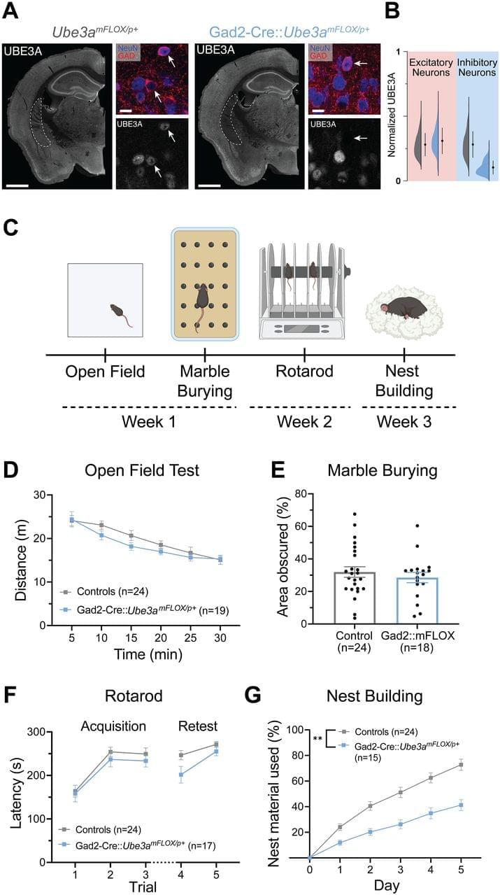
Our laboratory’s previous work identified an outsized role of GABAergic loss of UBE3A in hyperexcitability phenotypes. GABAergic loss of UBE3A drives increased delta power on cortical EEG (Judson et al., 2016), a phenotype that correlates with the severity of a range of symptoms in AS individuals (Hipp et al., 2021; Ostrowski et al., 2021). Further, mice with Ube3a deleted from GABAergic neurons show decreased threshold to chemically and acoustically driven seizures, and they also exhibit spontaneous behavioral seizures, a phenotype not observed in AS model mice on a C57BL/6J background (Judson et al., 2016; Gu et al., 2019). These data forewarn that UBE3A reinstatement in a manner biased to glutamatergic neurons could potentially worsen epilepsy-related symptoms and highlight the importance of studying the neuronal populations regulating other behaviors.

Based on the exaggerated role of GABAergic neurons in AS seizure phenotypes, we predicted that GABAergic deletion of Ube3a would underlie a broad range of behavioral phenotypes in AS mice. In the present study, we instead found a larger role of Ube3a deletion from glutamatergic neurons in motor coordination, measured by rotarod and open field behavior, and innate species-specific behaviors such as marble burying. Furthermore, glutamatergic loss of UBE3A appears to mediate alterations in sleep patterning and induces some sleep fragmentation, while UBE3A loss from GABAergic neurons only caused fragmented sleep. Interestingly, glutamatergic reinstatement of Ube3a also rescued the decreased REM sleep observed in AS mice, as estimated by the PiezoSleep system. While this study identified some roles of GABAergic neurons in nest building behavior and sleep fragmentation, our data largely suggest a divergence of the neural circuitry underlying the motor, innate behavior, and sleep phenotypes of AS mice from the circuitry responsible for seizure susceptibility and cortical EEG patterns.

/* */