Researchers discovered that <i>R. inulinivorans</i> plays an important role in muscle strength and could act as a probiotic candidate for nutraceutical interventions targeting age-related muscle-wasting diseases.


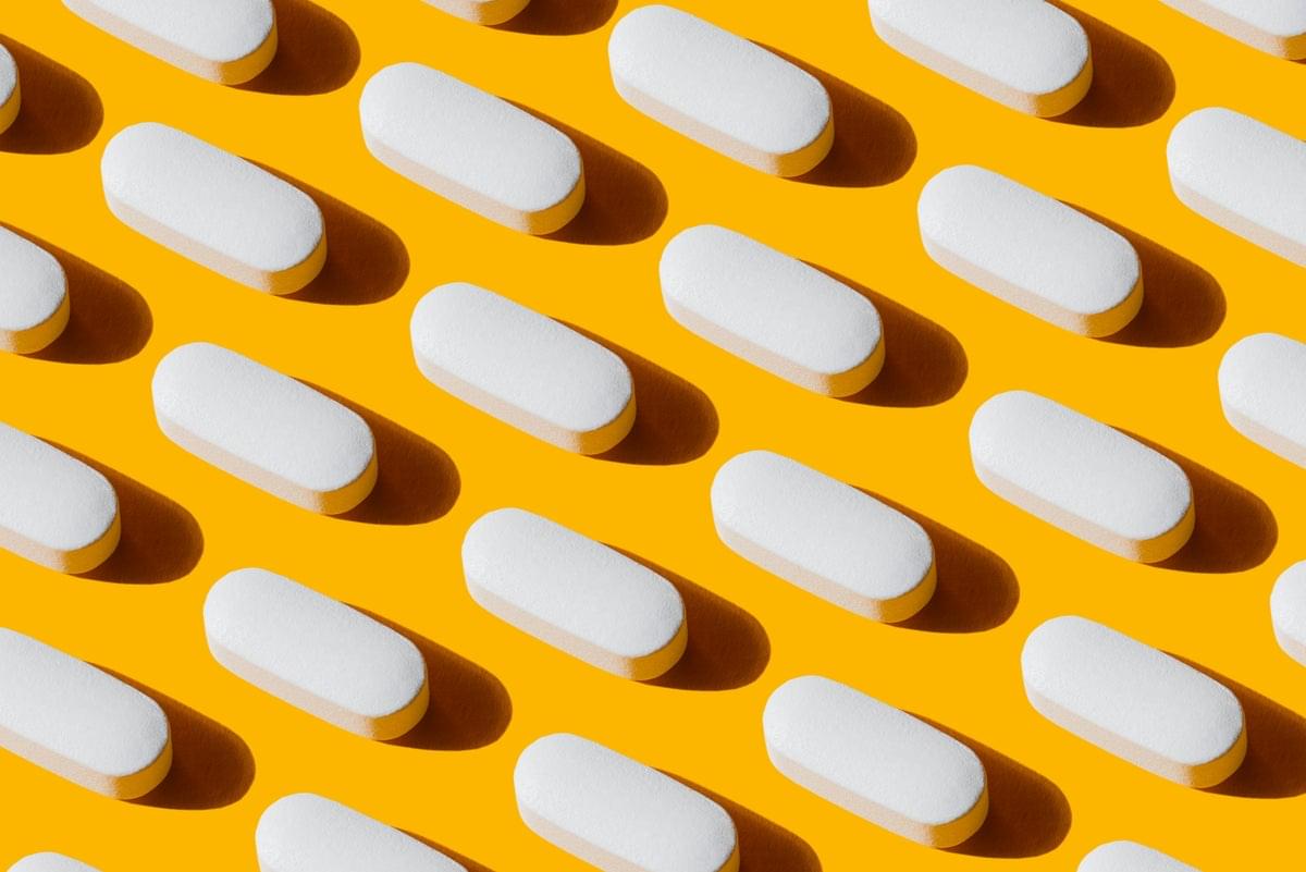
A research team from the University of Minnesota has discovered that certain polyunsaturated lipids (fatty acids) can selectively eliminate senescent cells — aged, dysfunctional cells that accumulate in the body over time and contribute to chronic disease and aging. The mechanism involves triggering ferroptosis, a regulated form of cell death, which senescent cells are particularly vulnerable to due to their elevated iron levels and heightened oxidative stress. This marks the first demonstration that fatty acids can act as senolytics (agents that clear senescent cells). While clinical application remains premature — further testing on animal models of age-related diseases is still needed — the findings open a promising new avenue for developing senolytic therapies targeting aging and its associated conditions.
MINNEAPOLIS/ST. PAUL (03/12/2026) —New research from the University of Minnesota Medical School has identified fatty acids that selectively induce death in senescent cells — the culprits behind aging and many chronic diseases, opening new avenues for age-related therapies. The findings were recently published in Cell Press Blue.
The research team discovered certain naturally occurring polyunsaturated lipids can selectively remove senescent cells. Senescent cells are old, damaged cells that accumulate with age and contribute to aging and many age-related diseases like pulmonary fibrosis, osteoarthritis and loss of resilience to infections.
These lipids cause senescent cells to die through a process called ferroptosis, which is a regulated form of cell death that occurs when iron in the cell triggers damaging reactions in its fats. The study also showed that these aging cells have high levels of iron and oxidative stress, which makes them uniquely susceptible to this process. Since lowering the number of senescent cells is associated with better health in old age, these natural, active fats could be used as a treatment for age-related illnesses caused by cellular senescence.

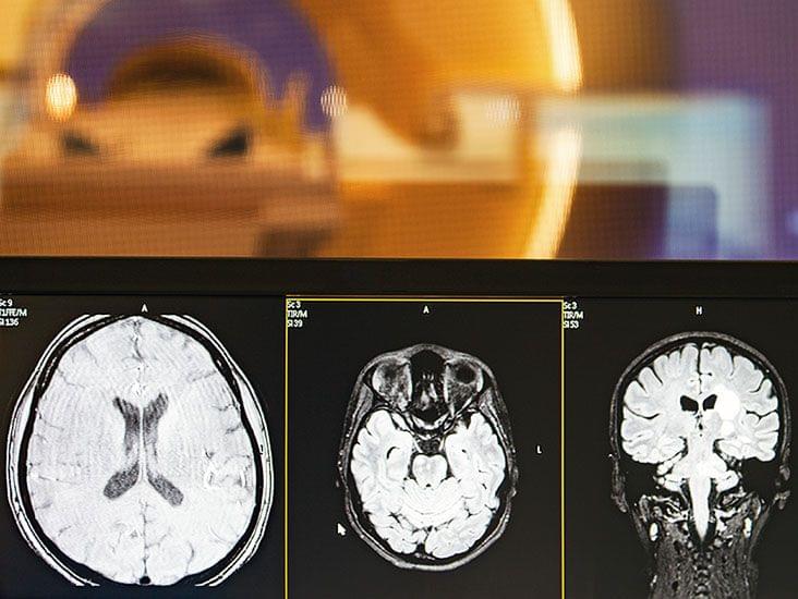
Scientists have plenty of ideas about why aging impairs memory. Reductions in blood flow in the brain, shrinking brain volume, and malfunctioning neural repair systems have all been blamed. Now, new research in mice points to another possible culprit: microbes in the gut.
In a new study, scientists show how a bacterium that is particularly common in older animals can drive memory loss. This microbe makes compounds that impair signaling along neurons connecting the gut with the brain, dampening activity in brain regions associated with learning and memory, the team found.
Research suggests the microbiome may contribute to cognitive decline—but its relevance in humans is unclear.
This actually offers some significant new insights for both cancer treatment research and the development of anti-aging therapies. 🧠
Read more👇
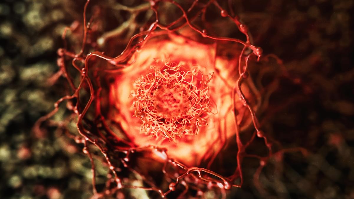
A group of natural compounds attracting attention for their anti-aging potential has a dark side.
New research shows how a family of chemicals called polyamines speeds up the growth of cancer cells. Led by a team from the Tokyo University of Science in Japan, the study offers some significant new insights for both cancer treatment research and the development of anti-aging therapies.
Polyamines are essential molecules found in all living cells. Including compounds with colorful names like spermidine and putrescine, they regulate processes involving cell growth and protein synthesis.
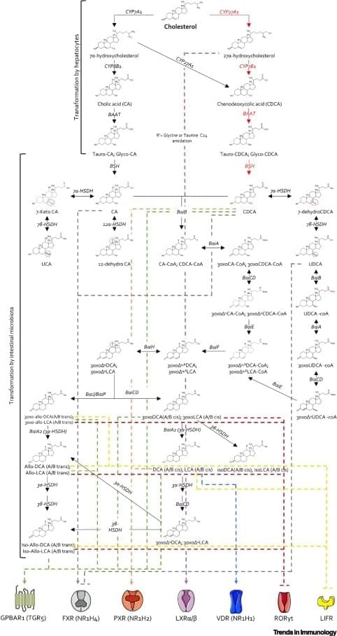
The human liver makes two primary bile acids that are cholesterol derivates, while, intestinal microbiota is the source of hundreds of secondary bile acids and microbially conjugated bile acids.
A dysbiotic microbiota releases altered quantities and varieties of secondary bile acids, which contribute to intestinal and systemic immune dysregulation.
In this review the authors discuss recent advances in secondary bile acids, the intestinal microbiota generating them, and their role in immune disorders. sciencenewshighlights ScienceMission https://sciencemission.com/Secondary-bile-acids
Bile acids are cholesterol derivatives, generated by the coordinated intervention of human and bacterial genes, functioning as endogenous ligands for multiple transcription factors and receptors throughout the body. While only two primary bile acids are generated by the human liver, the intestinal microbiota is the source of hundreds of secondary bile acids and microbially conjugated bile acids. Secondary bile acids regulate immune function throughout the body, promote the conversion of thyroid hormone, and regulate energy expenditure in muscle and adipose tissues, ultimately contributing to the beneficial effects of calorie restriction on human health and longevity. Here, we discuss recent advances in our understanding of secondary bile acids, the intestinal microbiota generating them, and their role in immune disorders.
For sexual reproduction to yield healthy offspring, newly generated oocytes—immature egg cells—must receive the correct amount of DNA after cell division. This process of segregating chromosomes becomes more prone to failure as we age. Now, RIKEN researchers have identified a strategy that could help to prevent such errors and restore healthy production of oocytes.
Oocytes are produced by a cell-division process known as meiosis, during which every chromosome is duplicated. These replicates form X-shaped structures in which the chromosomes are joined via structures called centromeres, where a protein called cohesin locks chromosome copies together.
As division proceeds, protein fibers called microtubules spread from opposite poles of the dividing cell, attaching to each chromosome. These microtubules eventually pull the two apart, so that each newly formed cell receives one copy of each chromosome.
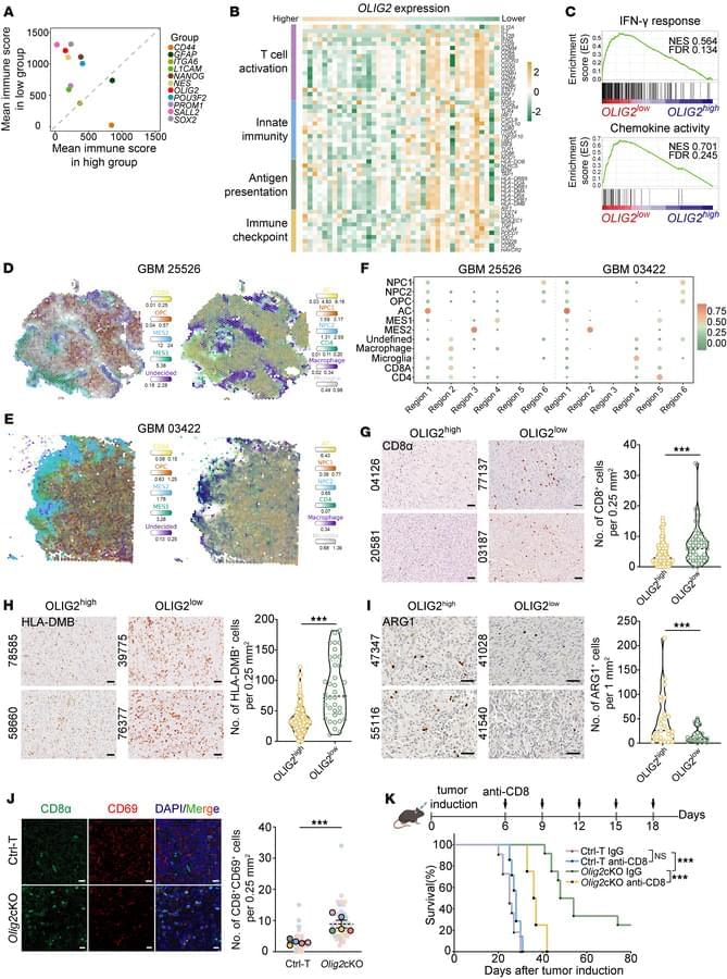
Here, Fanghui Lu & team report OLIG2, a master transcription factor in glioblastoma stem cells, enables immune evasion by suppressing CXCL10. And, targeting OLIG2 overcomes immunotherapy resistance and improves survival.
1Department of Cancer Center, The Second Affiliated Hospital of Chongqing Medical University, Chongqing, China.
2Department of Neurosurgery, Key Laboratory of Major Brain Disease and Aging Research (Ministry of Education), The First Affiliated Hospital of Chongqing Medical University, Chongqing, China.
3School of Basic Medical Sciences, Chongqing Medical University, Chongqing, China.