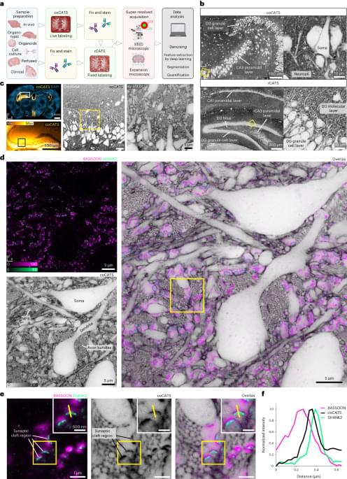
Another excellent paper from Johann G. Danzl’s research group. They develop methods that combine novel negative staining techniques, deep learning, and super-resolution STED microscopy or expansion microscopy to facilitate nanoscale-resolution imaging of brain tissue volumes. They also show semi-automated (and some fully automated) segmentation of neuron morphology and identification of synapses. Very cool work and I’m excited to see how it influences connectomics in the future! #brain #neuroscience #imaging #microscopy #neurotech
Mapping fixed brain samples with extracellular labeling and optical microscopy reveals synaptic connections.