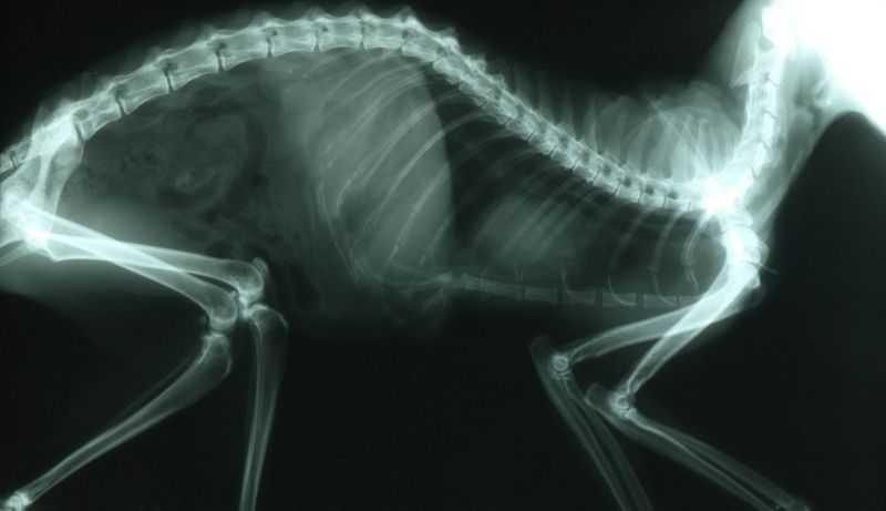
Scientist or not, we’re all familiar with X-ray imaging and perhaps its 3D cousin, computed tomography (CT), as well. These platforms are great for looking at bone and dense tissue—to see if there’s a fracture, or maybe a mass in the lung where it shouldn’t be—whereas molecular resonance imaging (MRI) and ultrasonography are the go-to modalities for interrogating softer tissue, like muscle. And for knowing what is happening in the body—as opposed to just where something is—nuclear tracer technologies like positron emission tomography (PET), and to a lesser extent its cousin single-photon emission computed tomography (SPECT), are the way to go.
These self-same modalities can be found in more diminutive instrumentation for pre-clinical imaging—often equipped with heated beds or chambers, anesthesia and oxygen supplies, and other modifications—specifically designed for small animals. If you also consider instruments capable of optical modalities of fluorescence, bioluminescence and their derivatives—which generally don’t easily translate to the clinic—you find yourself awash in possibilities for in vivo imaging.
