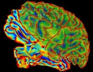
Two interconnected brain areas — the hippocampus and the entorhinal cortex — help us to know where we are and to remember it later. By studying these brain areas, researchers at Baylor College of Medicine, Rice University, The University of Texas MD Anderson Cancer Center and the National Cancer Institute have uncovered new information about how dysfunction of this circuit may contribute to memory loss in Alzheimer’s disease. Their results appear in Cell Reports.
“We created a new mouse model in which we showed that spatial memory decays when the entorhinal cortex is not functioning properly,” said co-corresponding author Dr. Joanna Jankowsky, associate professor of neuroscience at Baylor. “I think of the entorhinal area as a funnel. It takes information from other sensory cortices — the parts of the brain responsible for vision, hearing, smell, touch, and taste — and funnels it into the hippocampus. The hippocampus then binds this disparate information into a cohesive memory that can be reactivated in full by recalling only one part. But the hippocampus also plays a role in spatial navigation by telling us where we are in the world. These two functions converge in the same cells, and our study set out to examine this duality.”
The new mouse model was genetically engineered to carry a particular surface receptor on the cells of the entorhinal cortex. When this receptor was activated by administering the drug ivermectin to the mice, the cells of the entorhinal cortex silenced their activity. They stopped funnelling information to the hippocampus. This system allowed the scientists to turn off the entorhinal cortex, and to determine how this affected hippocampal function.
