And going forward, we’ll do this with far more knowledge of what we’re doing, and more control over the genes of our progeny. We can already screen ourselves and embryos for genetic diseases. We could potentially choose embryos for desirable genes, as we do with crops. Direct editing of the DNA of a human embryo has been proven to be possible — but seems morally abhorrent, effectively turning children into subjects of medical experimentation. And yet, if such technologies were proven safe, I could imagine a future where you’d be a bad parent not to give your children the best genes possible.
Computers also provide an entirely new selective pressure. As more and more matches are made on smartphones, we are delegating decisions about what the next generation looks like to computer algorithms, who recommend our potential matches. Digital code now helps choose what genetic code passed on to future generations, just like it shapes what you stream or buy online. This might sound like dark science fiction, but it’s already happening. Our genes are being curated by computer, just like our playlists. It’s hard to know where this leads, but I wonder if it’s entirely wise to turn over the future of our species to iPhones, the internet and the companies behind them.
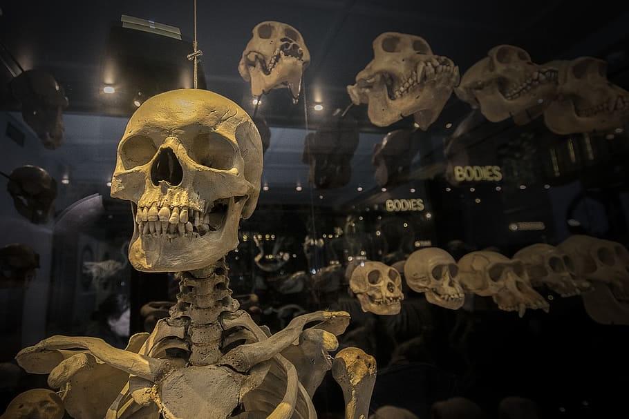
Discussions of human evolution are usually backward looking, as if the greatest triumphs and challenges were in the distant past. But as technology and culture enter a period of accelerating change, our genes will too. Arguably, the most interesting parts of evolution aren’t life’s origins, dinosaurs, or Neanderthals, but what’s happening right now, our present – and our future.