“There are no hard limits imposed by biology or by physics that says that we can’t live better longer,” Kristen Fortney, CEO of San Francisco-based BioAge Labs, told the outlet. Focused on discerning the markers of aging, BioAge Labs is using large amounts of biobank blood and tissue samples to do so.
The company has already found a drug target that slows aging-linked muscle loss in mice.
“There is a protein called apelin that circulates in the blood, and we saw that middle-aged people with higher levels of apelin in their blood were living longer, with better muscle function and better cognitive function as they age,” Fortney said, according to Express.
A new study in an animal model of aging indicates a potential reason for why women who have early menopause or other genetic conditions affecting the reproductive system are more prone to develop cardiovascular disease, diabetes, and dementia.
The new study, led by researchers from the University of Pittsburgh and UPMC and published in the journal Aging Cell, found that disrupting a process called meiosis in C. elegans reproductive cells caused a decline in the worms’ health and triggered an accelerated aging gene signature similar to that of aging humans.
“This study is exciting because it’s the first direct evidence that manipulating the health of reproductive cells leads to premature aging and a decline in healthspan,” said senior author Arjumand Ghazi, Ph.D., associate professor of pediatrics, developmental biology, and cell biology and physiology at Pitt and UPMC Children’s Hospital of Pittsburgh. “The implications of this finding are profound: It suggests that the status of the reproductive system is important not simply to produce children, but also for overall health.”
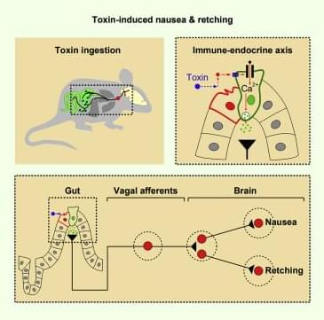
Identification of molecularly defined gut-to-brain and downstream brain circuits participating in nausea and retching induced by enterotoxins and chemotherapeutic drugs in mice suggests that food poisoning and chemotherapy recruit similar circuit modules to initiate defensive responses.
Thank you to Squarespace for sponsoring today’s video! Head to https://www.squarespace.com/anna to save 10% off your first purchase of a website or domain using code ANNA
According to an article by the university, the study is significant because it is a sign of future viability for noninvasive technology to help those with limited motor function.
“We demonstrated that the people who will actually be the end users of these types of devices are able to navigate in a natural environment with the assistance of a brain-machine interface,” said José del R. Millán, professor at the Cockrell School of Engineering’s Chandra Family Department of Electrical and Computer Engineering and leader of the international research team.
People affected by the lethal glioblastoma cancer only live for 12–18 months after diagnosis.
A global trial that began in 2007 has confirmed that a vaccine for the treatment of the most lethal brain cancer can give patients years of extended life.
Glioblastoma is not only the most common form of brain cancer but is also one of the deadliest. People affected by the disease only live just 12–18 months after the diagnosis, or even less.
Now, thanks to the vaccine DCVax, some 2,500 people who are diagnosed with deadly cancer in the UK could be benefitted.
Guarding Against Future Global Biological Risks — Dr. Margaret “Peggy” Hamburg, MD — Chair Nuclear Threat Initiative, bio Advisory Group; Commissioner, Bipartisan Commission on Biodefense; former Commissioner, U.S. Food and Drug Administration (FDA)
Dr. Margaret “Peggy” Hamburg, MD is an internationally recognized leader in public health and medicine, who currently serves as chair of the Nuclear Threat Initiative’s (NTI) bio Advisory Group (https://www.nti.org/about/people/margaret-hamburg-md/), where she has also served as founding vice president and senior scientist. She also currently holds a role as Commissioner on the Bipartisan Commission on Biodefense (https://biodefensecommission.org/teams/margaret-a-hamburg/).
Dr. Hamburg previously served as foreign secretary of the National Academy of Medicine and is a former Commissioner of the U.S. Food and Drug Administration (FDA), having served for almost six years where she was well known for advancing regulatory science, modernizing regulatory pathways, and globalizing the agency. Previous government positions include Assistant Secretary for Planning and Evaluation, U.S. Department of Health and Human Services, Health Commissioner for New York City, and Assistant Director of the National Institute of Allergy and Infectious Diseases, National Institutes of Health.
In her role, as Foreign Secretary of the National Academy of Medicine, the health arm of the National Academy of Sciences, Engineering and Medicine, Dr. Hamburg served as senior advisor on international matters and was the liaison with other Academies of Medicine around the world. She is an elected member of the Council on Foreign Relations and the National Academy of Medicine.
Dr. Hamburg currently sits on the boards of the Commonwealth Fund, the Simons Foundation, the Urban Institute, the Global Alliance for Vaccines and Immunization, the Parker Institute for Cancer Immunotherapy and the American Museum of Natural History. She is chair of the Joint Coordinating Group for the Coalition for Epidemic Preparedness and Innovation, and a member of the Harvard University Global Advisory Council, the Global Health Scientific Advisory Committee for the Gates Foundation, the Harvard Medical School Board of Fellows, and the World Dementia Council.
Current flowing through the plasma membrane of individual neurons causes fluctuations in the surrounding electrical field that can be detected with extracellular electrodes. Changes in the local field potential can influence the activity of all neurons within that field. For example, when two unmyelinated axons are closely apposed, an action potential in one axon alters the membrane potential of the other. This phenomenon is called ephaptic signaling. Ephaptic signaling is most prominent in layered neural structures in which numerous similarly oriented neurons are synchronously active. In fact, ephaptic signaling is thought to promote synchronous firing of cerebellar Purkinje cells and cortical and hippocampal pyramidal neurons. Ikeda et al. now show that ephaptic signaling originating in Drosophila eyes can influence activity in olfactory sensory neurons (OSNs) in the antennae.
It’s been seven years since Nigel French was woken up in the middle of the night by his wife after having a seizure, which came out of the blue after experiencing a mild headache – something he had simply put down to blocked sinuses.
“She told me that the ambulance had arrived and I was like: ‘what ambulance?’” recalls French, 53, a mechanic who was diagnosed with glioblastoma that required urgent surgery, without which he would have had only months to live.
In a different scenario, the impact of one of the most aggressive forms of brain cancer would have taken its toll by now, but thanks to a revolutionary vaccine, he is not only still alive but continuing to work and enjoy all that life has to offer.