Vasireddy et al. provide a framework for electrical stimulation current steering using several microelectrodes to most effectively target individual neurons in a population. A biophysically inspired mathematical model fits the linear and nonlinear responses of neurons, and data-driven regression models are used to efficiently find the most selective electrical stimulation patterns.
Are we thinking about consciousness in the wrong way?
Nick Lane is an multi-award-winning biochemist and an outstanding science communicator in the origins of life field. He is a Professor of Evolutionary Biochemistry at University College London.
Tap the link to watch his talk now.
How could calcium ions rushing through a membrane generate the taste of coffee, the smell of a rose or the feeling of love? Join celebrated biochemist, Nick Lane, as he argues that the deep logic of life is at root an electrical phenomenon.’His theories are ingenious, breathtaking in scope, and challenging in every sense’ — The Guardian.
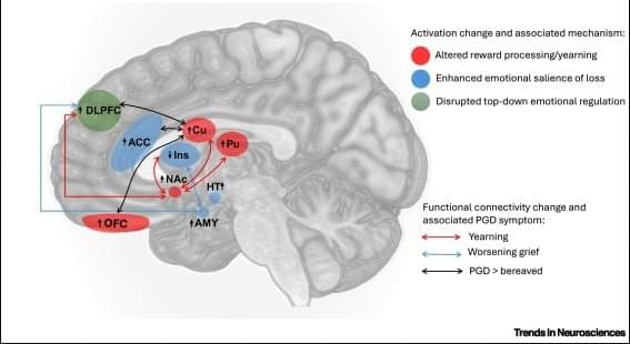
The neurobiology of why some brains cannot move on from loss.
Prolonged grief disorder (PGD) is a psychiatric condition that describes individuals who experience persistent grief reactions characterized by preoccupation with the loss. This review provides an overview of the evidence on neurobiological processes associated with PGD. We propose that, although the neurobiological circuitry of PGD overlaps with that of anxiety and depression, it also involves neural responses that reflect the distinct symptom profiles of people with PGD. Specifically, while recruitment of cognitive control and salience networks is observed across common mental disorders, there is evidence that aberrant neural processes implicated in reward processes and appetitive functions are somewhat distinctive to PGD.
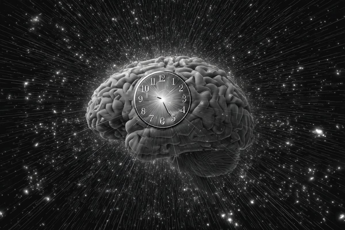
Forget everything you knew about practice making perfect. New research shows your brain is actually wired to learn faster from rare events than from constant repetition.
Structurally, they look similar: MNK1 and MNK2 belong to the same enzyme family and are best known for regulating how cells make proteins. Their starring role in such a crucial cellular function has cast them into the spotlight as potential drug targets to treat nervous system disorders and chronic pain. But would it matter whether a drug targets only one of them?
In a study published in Molecular Psychiatry, researchers led by Rosalba Olga Proce, a doctoral student in the Molecular and Cellular Basis of Behavior lab led by Dr. Hanna Hornberg at the Max Delbruck Center, set out to determine whether the two enzymes—also called kinases—perform distinct functions in the brain. The team found clear differences. Mice lacking MNK1 showed less interest in newly introduced objects than controls and impaired memory of objects. By contrast, mice without MNK2 appeared normal in object recognition tests but showed enhanced interest in social contacts.
“The behavioral differences we observed suggest that each kinase has a specialized function,” says Proce. “It might be preferable to target each kinase individually when designing drugs.”
Is there something that it is like to be an electron? That sounds implausible. Yet Galen Strawson believes this is the best explanation of how things are.
Specifically, Galen offers his view on physicalistic panpsychism (though there are non-physicalistic panpsychisms as well). He argues something like this, it seems to me:
First, Galen assumes (very plausibly) that experiential phenomena are real phenomena, opposed to illusory. Now:
1. If radical emergentism is true, then experiential phenomena emerges from wholly and utterly non-experiential phenomena. 2. But experiential phenomena cannot emerge from wholly and utterly non-experiential pheneomena. 3. So radical emergentism is false. [1, 2] 4. If radical emergentism is false, then experiential phenomena must already exist in some sense and to some extent as a feature of physical stuff to give rise to experiential phenomena in an intelligible way. 5. So experiential phenomena must already exist in some sense and to some extent as a feature of physical stuff to give rise to experiential phenomena in an intelligible way. [3, 4]
In other words, consciousness has been a feature of the universe since the Big Bang.
For sustained and scholarly treatment of panpsychism, see Galen Strawson’s paper, \.
More than a century ago, Pavlov trained his dog to associate the sound of a bell with food. Ever since, scientists have assumed the dog learned this through repetition. The more times the dog heard the bell and then got fed, the better it learned that the sound meant food would soon follow.
Now, scientists at UC San Francisco are upending this 100-year-old assumption about associative learning. The new theory asserts that it depends less on how many times something happens and more on how much time passes between rewards.
“It turns out that the time between these cue-reward pairings helps the brain determine how much to learn from that experience,” said Vijay Mohan K. Namboobidiri, Ph.D., an associate professor of Neurology and senior author of the study, published in Nature Neuroscience.
Benfey et al. find that norepinephrine shifts the visual response selectivity of optic tectal neurons in the Xenopus tadpole to favor threatening loom stimuli over more neutral, randomly drifting dots. Mechanistically, norepinephrine induces radial astrocyte activation and glial release of ATP/adenosine, resulting in reduced excitatory neurotransmission and selectivity shift.