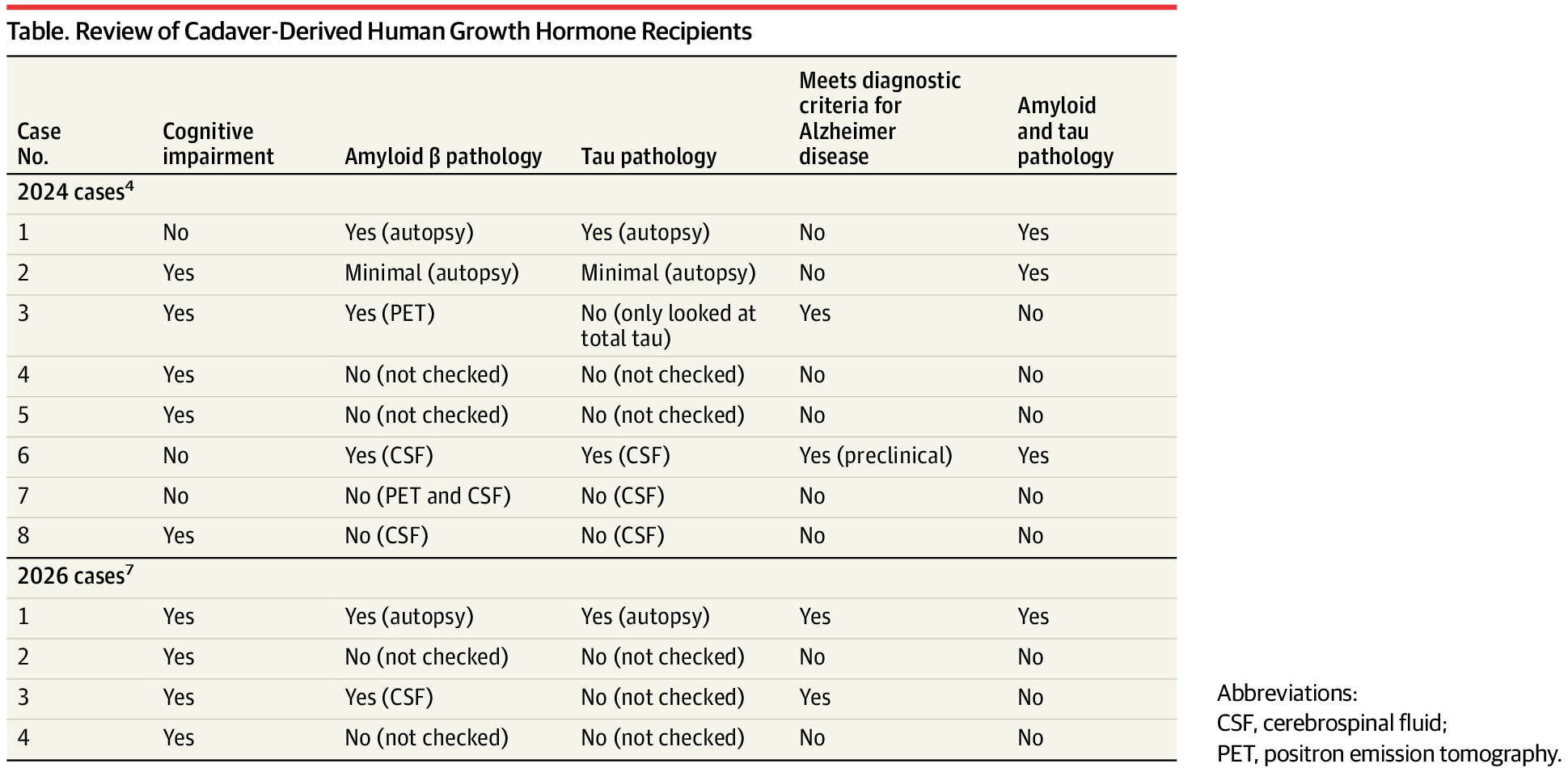
Toggle light / dark theme

In today’s interconnected world, infectious diseases pose an escalating threat, as demonstrated by the coronavirus pandemic and outbreaks of H1N1, SARS, Ebola, Zika, and H5N1 (bird flu) viruses—all of which have had significant global health and economic impacts.

But more common viral diseases also contribute to global health challenges and economic costs. For example, seasonal influenza epidemics occur annually, causing a substantial global disease burden and economic losses exceeding $11.2 billion each year in the United States alone. Meanwhile, herpes simplex virus-1 (HSV-1), spread primarily through oral contact, infects over two-thirds of the global population and is the leading cause of infectious blindness in Western countries.

Low vaccination rates for influenza viruses and the lack of an HSV vaccine underscore the need for a new approach—one that targets reducing viral loads at the sites where transmission occurs. And for viruses like these, which are transmitted more efficiently through the mouth than the nose, this means focusing on the oral cavity.

Should you step away from the chicken wings?

For years, the conventional wisdom has been to swap out red meat for white meats like chicken and poultry to help reduce health risks like increased cholesterol, cancer, and inflammation —not to mention get a more budget-friendly protein source. But a new study links eating chicken and other poultry with a significantly increased risk of dying from gastrointestinal cancer and all other causes.

But before you put down the chicken—or roll your eyes and get back to your chicken Caesar—check out the details of the study, and what a dietitian says you should do if you’re concerned.

The thymus is a crucial training ground for T-cells, the body’s “white knights,” where they learn to battle the various diseases they may encounter. Thymic function shrinks to nearly nothing as we age, severely limiting our ability to recognize and defend against cellular infiltrators.

Scientists at the University of Texas Health Science Center at San Antonio (UT Health San Antonio) discovered a crucial pathway in the thymus that determines the rate of growth and functional preservation. Surprisingly, this pathway appears to act through both indirect and direct methods. Understanding these functions could help produce treatments that preserve thymic function for longer, boosting the immune system’s power to fight disease.

A UT Health San Antonio-led study, published in Nature Aging in February 2025, highlights the role of the peptide hormone fibroblast growth factor 21 (FGF21) in regulating T-cells and, potentially, preserving thymic size over time.

Alternative RNA splicing is like a movie editor cutting and rearranging scenes from the same footage to create different versions of a film. By selecting which scenes to keep and which to leave out, the editor can produce a drama, a comedy, or even a thriller—all from the same raw material. Similarly, cells splice RNA in different ways to produce a variety of proteins from a single gene, fine-tuning their function based on need. However, when cancer rewrites the script, this process goes awry, fueling tumor growth and survival.

In a recent study reported in the Feb. 15 issue of Nature Communications, scientists from The Jackson Laboratory (JAX) and UConn Health not only show how cancer hijacks this tightly regulated splicing and rearranging of RNA but also introduce a potential therapeutic strategy that could slow or even shrink aggressive and hard-to-treat tumors. This discovery could transform how we treat aggressive cancers, such as and certain , where current treatment options are limited.

At the heart of this work, led by Olga Anczuków, an associate professor at JAX and co-program leader at the NCI-designated JAX Cancer Center, are tiny genetic elements called poison exons, nature’s own “off switch” for protein production. When these exons are included in an RNA message, they trigger its destruction before a protein can be made—preventing harmful cellular activity. In , poison exons regulate the levels of key proteins, keeping the genetic machinery in check. But in cancer, this safety mechanism often fails.

Over the past decades, electronics engineers developed increasingly small, flexible and sophisticated sensors that can pick up a wide range of signals, ranging from human motions to heartrate and other biological signals. These sensors have in turn enabled the development of new electronics, including smartwatches, biomedical devices that can help monitor the health of users over time and other wearable or implantable systems.

Strain , which are designed to convert mechanical force into , are among the most widely used sensing devices within the , as they can be valuable for tracking both human movements and health-related biological signals. While these sensors are already embedded in many electronic devices, most existing solutions are only able to track movements in one direction.

Sensors that can accurately pick up movements and forces in multiple directions could be highly advantageous, as they could be applied to a wider range of scenarios. In addition, these sensors could be embedded in existing electronic devices to broaden their functions or enhance their capabilities.

But classic risk factors do not seem to fully explain the recent rise in early-onset cancers, says Dr. Cathy Eng, director of the Young Adult Cancers Program at Vanderbilt University’s Ingram Cancer Center in Tennessee. Some of the trends are baffling; young, nonsmoking women, for example, are being diagnosed with lung cancer in strangely high numbers. Many times, Eng’s patients were extremely healthy: vegetarians, marathon runners, avid swimmers. “That’s why I really believe there’s other risk factors to account for this,” she says.

There’s no shortage of theories about what those may be. Many scientists point to modern diets, which tend to be heavy on potentially carcinogenic products—including ultra-processed foods, red meat, and alcohol—and may also contribute to weight gain, another cancer risk factor. The foods we eat can also affect the gut microbiome, the colony of microbes that lives in the digestive system and appears linked to overall health. Alterations to the gut microbiome via diet, or perhaps exposure to drugs like antibiotics, have also been implicated.

Other researchers blame the microplastics littering our environment and leaching into our food and water supplies, some of which, according to a 2024 study, have even shown up in cancer patients’ tumors. Other environmental factors could also be to blame, given that everything from cosmetics to food packaging contains substances that many researchers aren’t convinced are safe. Even our near constant exposure to artificial light could be messing with normal biological rhythms in ways that have profound health consequences, some research suggests.

The U.S. government is preparing to make moves to get food dyes out of what we eat– a plan which may spark curiosity across the nation as to what the potential health risks of artificial food dyes are.

The metabolic fitness of microglia is markedly impaired in TREM2 knockout (KO) models [58]. TREM2, through its adaptors DAP12 and DAP10, activates the mechanistic target of rapamycin (mTOR) signaling pathway, which plays a crucial role in regulating metabolic pathways and protein synthesis [11, 58]. Loss of TREM2 impairs mTOR activation, leading to reduced ATP production and biosynthesis. In vivo FDG-PET imaging of TREM2 KO and TREM2 T66M knock-in mice shows a significant reduction in cerebral glucose metabolism [67, 68]. This decrease may correlate with impaired glucose uptake by microglia. Supporting this, ex vivo measurements of isolated microglia from TREM2 KO animals reveal lower FDG uptake [68].

Given the pivotal role of microglial metabolism in AD, targeting this process represents a promising therapeutic strategy. Agents such as interferon-γ (IFN-γ) and cyclocreatine, which enhance ATP production, have been shown to restore microglial functions and mitigate AD pathology [58, 65]. Notably, TREM2-activating antibodies boost microglial energy metabolism by promoting mitochondrial fatty acid and glucose oxidation [69]. Moreover, translocator protein (TSPO)-PET and FDG-PET imaging have demonstrated that TREM2 activation enhances microglial activity and glucose metabolism in amyloid mouse models. Thus, targeting TREM2 and microglial metabolism may complement existing AD therapies, which primarily focus on amyloid clearance and synaptic dysfunction, providing a more comprehensive approach to disease intervention.

Lipid metabolism is crucial for maintaining microglial functions and CNS homeostasis, influencing cellular membrane integrity, energy storage, and inflammatory responses. Emerging evidence identifies TREM2 as a key regulator of lipid metabolism in the brain. TREM2 binds a diverse range of lipids, including anionic and zwitterionic species such as sphingomyelin, phosphatidic acid, phosphatidylinositol, phosphatidylcholine, phosphatidylglycerol, phosphatidylserine (PtdSer) and sulfatide [49, 53, 70]. Among these, PtdSer is the most abundant negatively charged phospholipid in the inner leaflet of the plasma membrane in eukaryotic cells [71]. In neurodegenerative conditions, PtdSer externalization on damaged or apoptotic neurons serves as an “eat-me” signal, triggering TREM2-dependent microglial synaptic pruning and cell clearance [72]. Super-resolution microscopy and in vivo imaging studies have demonstrated that Aβ oligomer-induced hyperactive synapses expose PtdSer, marking them for TREM2-mediated engulfment, which helps mitigate neuronal hyperactivity in AD models. Additionally, individuals carrying TREM2 loss-of-function variants exhibit an accumulation of apoptotic-like synapses [72], underscoring TREM2’s essential role in synaptic homeostasis during early AD pathology. Beyond synaptic pruning, TREM2 facilitates the recognition and clearance of damaged cells. Notably, over-expression of TREM2 in non-phagocytic cells, such as Chinese hamster ovary (CHO) and HEK293 cells, enables them to engulf apoptotic neurons, highlighting TREM2’s function in lipid sensing and phagocytosis [16, 73]. This broad lipid-binding capability underscores TREM2’s critical role in modulating microglial responses to neurodegenerative insults and preserving neuronal health.

Saying one thing while feeling another is part of being human, but bottling up emotions can have serious psychological consequences, such as anxiety or panic attacks. To help health care providers tell the difference, a team led by scientists at Penn State has created a stretchable, rechargeable sticker that can detect real emotions—by measuring things like skin temperature and heart rate—even when users put on a brave face.

The researchers recently unveiled the wearable patch that can simultaneously and accurately track multiple emotional signals in a study published in the journal Nano Letters.

“This is a new and improved way to understand our emotions by looking at multiple body signals at once,” said Huanyu “Larry” Cheng, the James L. Henderson, Jr. Memorial Associate Professor of Engineering Science and Mechanics at Penn State and lead author of the paper.