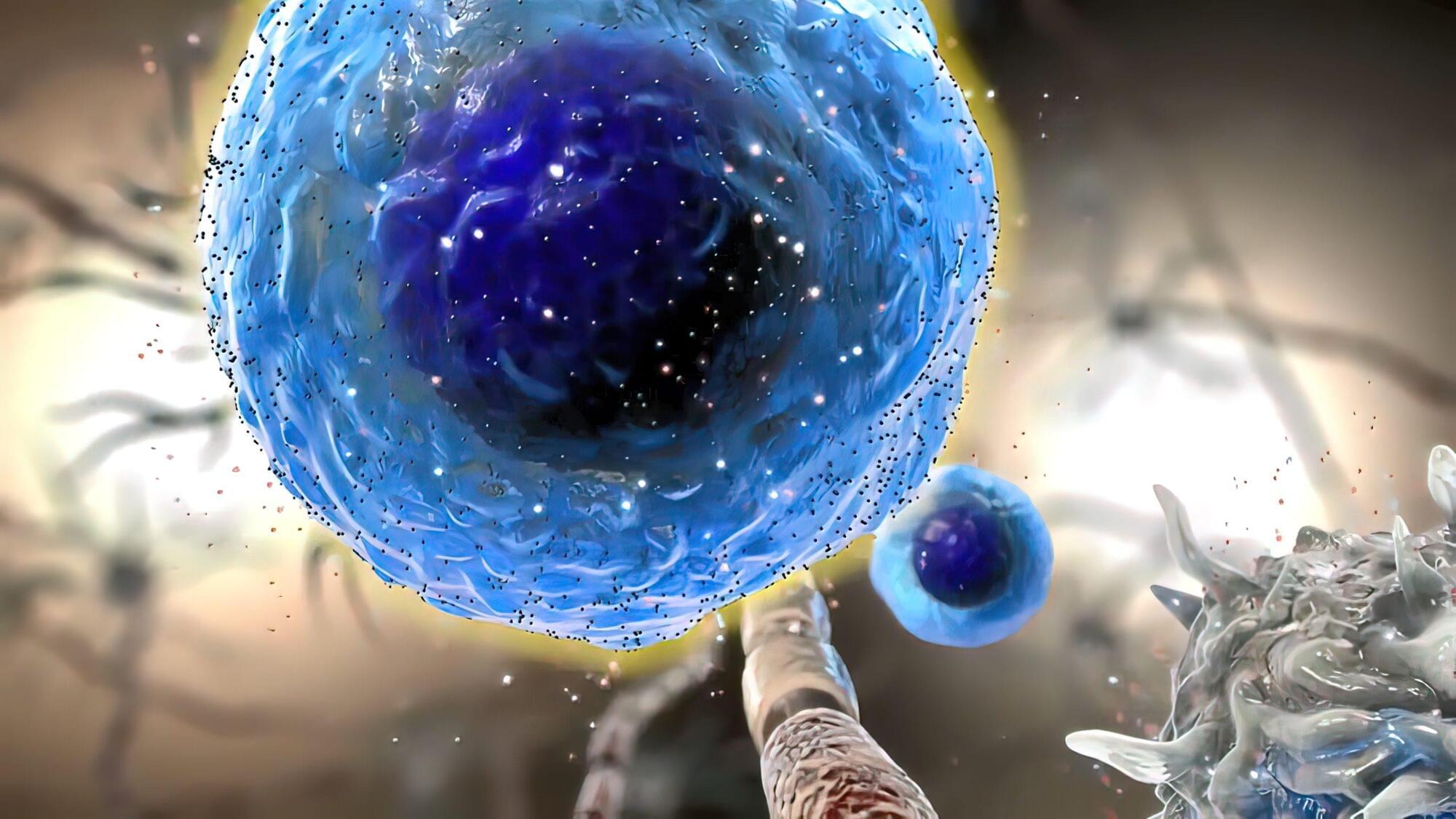
A UCLA study found that macrophages rely on interferon gamma signaling from past infections to preserve innate defenses.
What do you think about the foregoing arguments?
Interview from the Conference “Emergence and Panpsychism” in Munich 2011.
More information and the complete list of videos here: http://www.geiststaub.de/
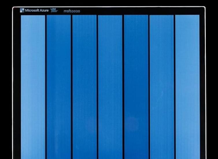
Scientists at Microsoft Research in the United States have demonstrated a system called Silica for writing and reading information in ordinary pieces of glass which can store two million books’ worth of data in a thin, palm-sized square.
In a paper published today in Nature, the researchers say their tests suggest the data will be readable for more than 10,000 years.
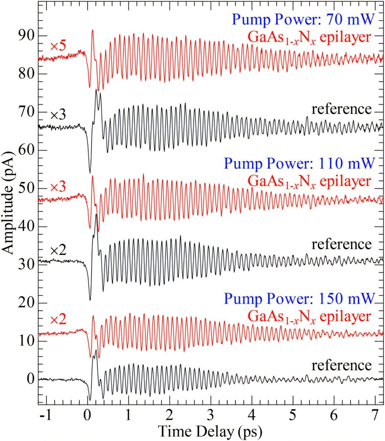
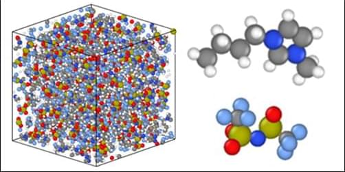
An Osaka Metropolitan University-led research team investigated the decay time of coherent longitudinal optical (LO) phonons both in a GaAs1−x Nx epilayer and in a GaAs single crystal to clarify the effects of dilute nitridation.
The team observed in terahertz time-domain spectroscopy that the terahertz electromagnetic waves, which are emitted from the coherent GaAs-like LO phonons, have a relatively long decay time in a GaAs1−x Nx epilayer in comparison with the terahertz waves from the coherent GaAs LO phonons in a semi-insulating GaAs single crystal.
This implies that alloy effects (mixed crystal effects) on the phonon Raman band broadening, which have a possibility of leading to the short decay time, hardly govern the decay time even in the present GaAs1−x Nx epilayer sample.

