Toggle light / dark theme

Genetic sequencing has aided in the discovery of novel mutations in inborn errors of immunity, but their clinical relevance often remains unclear

In this Research Article, Marcus Altfeld & team identify a TLR8 gene mutation causing immune overactivation and inflammation in two siblings, linking genetic change to immune system dysfunction and disease: Immunology.


14 Hamburg Center for Translational Immunology;

15 German Center for Child and Adolescent Health (DZKJ), partner site Hamburg; and.

16 Institute of Medical Microbiology, Virology and Hygiene, University Medical Center Hamburg-Eppendorf, Hamburg, Germany.

Cell-free DNA offers early warning for bloodstream infections in kids with leukemia

Researchers have identified a promising way to predict bloodstream infections in children with high-risk leukemia days before the infection would be diagnosed using current standards of care. The test, named plasma microbial cell-free DNA sequencing (mcfDNA-Seq), detected infection-causing pathogens days before standard blood cultures, offering a potential approach to protecting vulnerable patients by allowing treatment to start before the patient gets sick. The study is published in The Lancet Microbe.

Bloodstream infections are a major threat to children undergoing treatment for leukemia. Chemotherapy weakens the immune system, making even common bacteria or fungi potentially life-threatening. These infections can quickly lead to sepsis, prolonged hospital stays, delayed chemotherapy and, in some cases, early mortality. Catching these infections early is critical, yet clinicians currently have no reliable method to identify infections before symptoms appear.

“We’re not good at predicting or preventing infections in children with cancer, and the consequences can be deadly, causing lasting damage or delaying chemotherapy, which reduces the chances of successful treatment,” said first and corresponding author Joshua Wolf, Ph.D., MBBS, St. Jude Department of Infectious Diseases. “Many infections still happen even with the best prevention strategies we have, so what we really need is a way to detect infections before they start, so we can treat kids earlier and save lives.”

Insufficient Sleep in Adolescents and the Need for System Changes

💬 Editor’s Note by JAMA Editorial Fellow Randi Bates and JAMA Deputy Editor Tracy Lieu: Adolescents face increasing rates of insufficient sleep, driven by early school start times and digital media use, undermining cognitive and mental health.


Insufficient sleep is one of the most common health risks in adolescents and is associated with worse cognitive performance and academic achievement, as well as depression, other mental health conditions, and physical concerns. The American Academy of Sleep Medicine and American Academy of Pediatrics (AAP) recommend adolescents aged 13 to 18 years sleep for 8 to 10 hours each night.1 Yet, studies have found that adequate sleep eludes most adolescents.2

In this issue of JAMA, Bommersbach and colleagues report worsening trends in insufficient sleep duration among US high school students based on an analysis of the national Youth Risk Behavior Survey.3 Insufficient sleep increased from 68.9% in 2007 to 76.8% in 2023, largely from increases in very short sleep durations of less than or equal to 5 hours per night. This trend was observed in all demographic groups and was generally consistent across subgroups characterized by behavioral risk factors.

These sweeping patterns suggest that structural and environmental factors may be driving increases in insufficient sleep at the population level. Although some studies have focused on changing individual behavior to increase sleep, such interventions may have limitations in adolescents whose self-regulatory and decision-making abilities are still maturing. Additionally, adolescents may lack sufficient agency to overcome school or social system barriers that limit sleep.

Structures of hantavirus glycoprotein tetramers

This surface protein complex for the Andes virus is a mushroom-shaped structure called a Gn-Gc tetramer. To map the 3D structures, the team first produced virus-like particles that mimic a real virus, but without the genome that makes a virus infectious. They then used a cryo-electron microscope—which shines an electron beam through a frozen sample and detects the shadows created by molecules—to reconstruct the three-dimensional structures of the Gn-Gc tetramers on the surface of the virus-like particles.

But there was a twist: To obtain extremely high-resolution structures, the researchers painstakingly identified and isolated shadows from only the tetramers that were pointing sidewise relative to the electron beam and ignored those pointing in other directions. This allowed them to borrow a reconstruction method typically used on individual proteins.

The resulting structures have an extremely high resolution of 2.3 angstroms, meaning details the size of just a couple of atoms were effectively captured. That’s enough to represent a transformational improvement over another team’s model of the tetramer from a few years ago, at a resolution of 12 angstroms, still tiny but large enough to produce some key inaccuracies – ones effectively corrected with the newer method and resulting structure.

These latest structures show the Gn-Gc tetramer in a particular state before it has infected a cell. For vaccines or antibody therapies to be most effective against a hantavirus, mimicking surface proteins at this pre-infection stage is essential. ScienceMission sciencenewshighlights.


Hantaviruses, transmitted from rodents to people, have a death rate approaching 40%. They’re found around the world, and because there are no approved vaccines or treatments, they’re among the pathogens of highest concern for future pandemics. They made news in the United States last year when Betsy Arakawa, the wife of actor Gene Hackman, died from a hantavirus infection in New Mexico in March.

New findings published in the journal Cell about the Andes virus, a hantavirus endemic to the southwestern U.S. and other parts of North and South America, represent a crucial first step towards much-needed vaccines and antibody therapies for this and other hantaviruses.

A new class of Alzheimer’s biomarkers: Why protein shape may beat protein levels

Researchers have identified a new type of blood-based biomarker test for Alzheimer’s disease that measures structural changes in proteins, providing more information on the underlying biology of the disease than standard blood tests. The findings, published in Nature Aging, also provide new insights into how Alzheimer’s disease biology may differ between males and females.

“This work introduces a fundamentally new, blood-based approach to detecting and staging Alzheimer’s disease,” said Dr. Richard Hodes, director of NIH’s National Institute on Aging (NIA). “By revealing protein structural changes associated with genetic risk, symptom severity, and sex differences—features not captured by existing biomarkers—this research could enable earlier diagnosis and more effective clinical trials.”

A biotech company just doubled the lifespan of mice without changing their diet and without editing their genes

Instead, they trained the immune system to hunt down and destroy the cells that make the body age. Then they flooded the body with fresh stem cells to rebuild what was lost.

This isn’t science fiction. It’s longevity science happening right now.

Read more

Genomic reorganization at the transition to gametogenesis

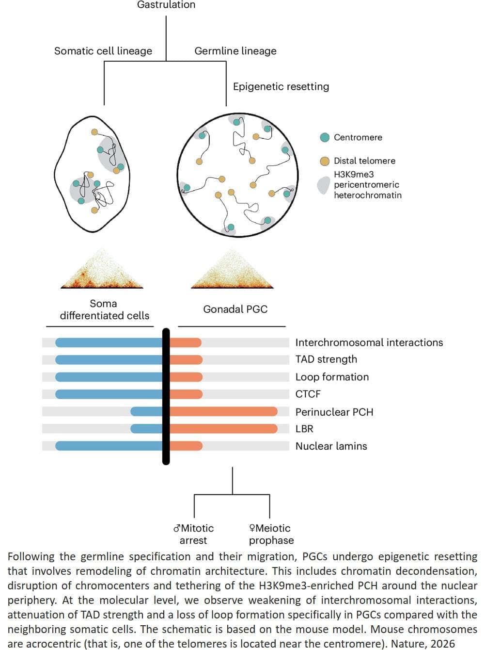
Using a technique called Hi-C analysis, which looks at how DNA is arranged in three dimensions inside the nucleus, the team found that at this transitional point the genome’s three-dimensional organisation becomes less structured and chromosomes become more separated inside the nucleus.

Creating sperm and eggs in the laboratory (in vitro) remains one of the greatest challenges in reproductive biology. To study this process, scientists use primordial germ cell–like cells (PGCLCs), which are lab-generated cells derived from embryonic stem cells that mimic the embryo’s earliest reproductive cells. However, these PCGLCs often fail to complete all the steps of meiosis, making it difficult to create functional sperm and eggs in petri dishes.

After studying the process in germ cells from the embryos, the team studied lab-generated mouse PCGLCs to see if the centromeres migrated to the periphery of the nucleus in vitro too, but they did not see the same phenomenon.

“The presence of this chromosome conformation in embryonic germ cells, but not lab-grown cells, suggests that this structural change could be required for meiosis to proceed properly, and could explain why meiosis is so difficult to recreate outside the body,” says the author, “but we need to do more work to fully characterise the process before we can say for sure.”

“Our study has uncovered a previously unknown and frankly very surprising restructuring of genome architecture that occurs in developing germ cells, which we believe is critical for a successful execution of meiosis,” says the senior author. ScienceMission sciencenewshighlights.


In our cells, our DNA carries chemical or ‘epigenetic’ marks that decide how genes will be used in different tissues. Yet in the group of specialised cells, known as ‘germ cells’, which will later form sperm and eggs, these inherited chemical instructions must be erased or reshuffled so development can begin again with a fresh blueprint in future generations.

Foundation AI model uses MRI data to predict multiple brain disorders

Artificial intelligence (AI) systems are computational models that can learn to identify patterns in data, make accurate predictions or generate content (e.g., texts, images, videos or sound recordings). These models can reliably complete various tasks and are now also used to carry out research rooted in different fields.

Over the past few decades, some AI models have proved promising for the early diagnosis and study of specific diseases or neuropsychiatric conditions. For instance, by analyzing large amounts of brain scans collected using a noninvasive technique known as magnetic resonance imaging (MRI), AI could uncover patterns associated with tumors, strokes and neurodegenerative diseases, which could help to diagnose these conditions.

Researchers at Mass General Brigham, Harvard Medical School and other institutes recently developed Brain Imaging Adaptive Core (BrainIAC), a large AI system pre-trained on a vast pool of MRI data that could be adapted to tackle different tasks. This foundation model, presented in a paper published in Nature Neuroscience, was found to outperform many models that were trained to complete specific medical or neuroscience-related tasks.

Protein Folding and Quality Control in the Endoplasmic Reticulum: Recent Lessons from Yeast and Mammalian Cell Systems

The evolution of eukaryotes was accompanied by an increased need for intracellular communication and cellular specialization. Thus, a more complex collection of secreted and membrane proteins had to be synthesized, modified, and folded. The endoplasmic reticulum (ER) thereby became equipped with devoted enzymes and associated factors that both catalyze the production of secreted proteins and remove damaged proteins. A means to modify ER function to accommodate and destroy misfolded proteins also evolved. Not surprisingly, a growing number of human diseases are linked to various facets of ER function. Each of these topics will be discussed in this article, with an emphasis on recent reports in the literature that employed diverse models.

/* */