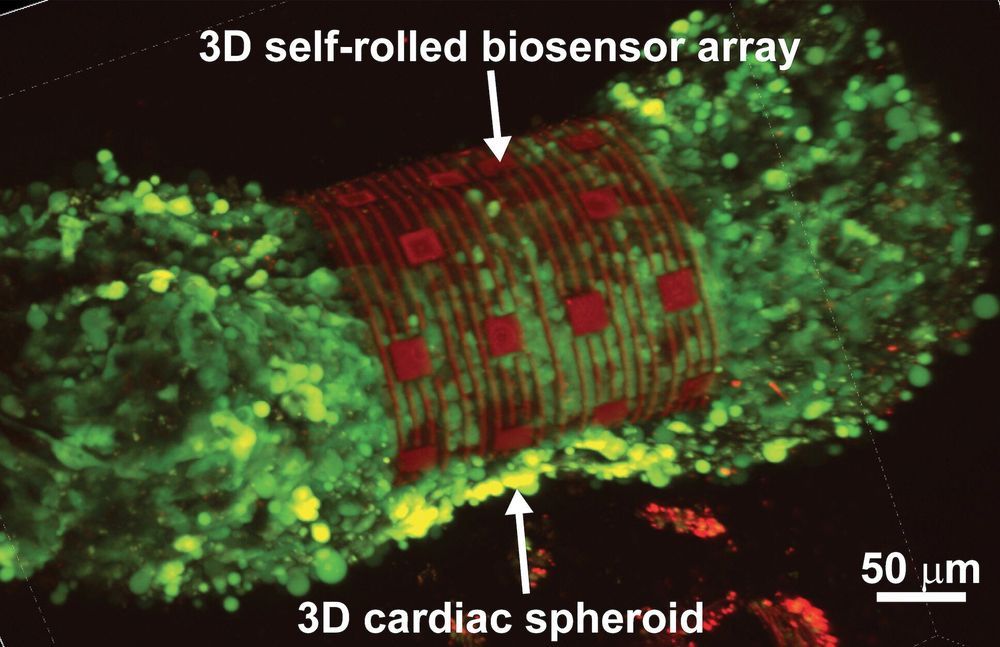
Researchers from Carnegie Mellon University (CMU) and Nanyang Technological University, Singapore (NTU Singapore) have developed an organ-on-an-electronic-chip platform, which uses bioelectrical sensors to measure the electrophysiology of the heart cells in three dimensions. These 3D, self-rolling biosensor arrays coil up over heart cell spheroid tissues to form an “organ-on-e-chip,” thus enabling the researchers to study how cells communicate with each other in multicellular systems such as the heart.
The organ-on-e-chip approach will help develop and assess the efficacy of drugs for disease treatment—perhaps even enabling researchers to screen for drugs and toxins directly on a human-like tissue, rather than testing on animal tissue. The platform will also be used to shed light on the connection between the heart’s electrical signals and disease, such as arrhythmias. The research, published in Science Advances, allows the researchers to investigate processes in cultured cells that currently are not accessible, such as tissue development and cell maturation.
“For decades, electrophysiology was done using cells and cultures on two-dimensional surfaces, such as culture dishes,” says Associate Professor of Biomedical Engineering (BME) and Materials Science & Engineering (MSE) Tzahi Cohen-Karni. “We are trying to circumvent the challenge of reading the heart’s electrical patterns in 3D by developing a way to shrink-wrap sensors around heart cells and extracting electrophysiological information from this tissue.”
