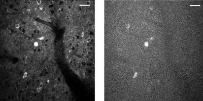
A three-photon microscopic video of neurons in a mouse brain. The imaging depth is approximately 1 millimeter below the surface of the brain. The firing of the neurons is captured by an indicator that is based on green fluorescent protein GFP, which glows brighter as the neuron sends a signal.
Nearly four years ago, then-President Obama launched the BRAIN (Brain Research through Advancing Innovative Neurotechnologies) Initiative, to “accelerate the development and application of new technologies that will enable researchers to produce dynamic pictures of the brain.”
Several of the program’s initial funding awards went to Cornell’s Chris Xu, the Mong Family Foundation Director of Cornell Neurotech – Engineering, and professor and director of undergraduate studies in applied and engineering physics. Xu’s projects aimed to develop new imaging techniques to achieve large scale, noninvasive imaging of neuronal activity.
